
Aleph Neuro has unveiled high-resolution three-dimensional vascular images of a living human brain captured through the intact skull using a system built with Butterfly Network’s Ultrasound-on-Chip platform. The research-stage result uses contrast-enhanced ultrasound localization microscopy and gives Butterfly Network’s embedded technology business a potentially important neuroimaging use case, although the work has not yet established clinical utility, regulatory readiness or broad reproducibility.
Why the Aleph Neuro images matter more as a vascular imaging advance than a finished brain interface
The most important distinction is that Aleph Neuro has demonstrated detailed imaging of cerebral vasculature, not direct reading of neurons, thoughts or cognitive content. The technique follows microbubbles moving through blood vessels and reconstructs their positions over time, producing a highly detailed map of vascular structure and flow. That is scientifically relevant because cerebral blood delivery is closely linked to neural activity, but the relationship is indirect and should not be confused with electrophysiological recording.
This distinction matters because the language surrounding brain-computer interfaces can quickly outrun the evidence. Aleph Neuro’s long-term ambition is to develop a non-invasive interface capable of accessing distributed brain activity without implanted electrodes or a room-sized scanner. The newly disclosed images provide an enabling hardware and imaging milestone, but they do not yet show reliable decoding of speech, movement intention, memory or perception in humans.
The immediate value may therefore lie in neurovascular imaging rather than consumer-facing brain interfaces. Stroke, traumatic brain injury, neurodegenerative disease and other neurological conditions can alter blood flow, vessel structure or microvascular function. A non-ionising system capable of visualising those changes at high resolution could eventually support research, diagnosis or treatment monitoring, but each indication would require its own evidence base, clinical endpoint and regulatory strategy.
How ultrasound localization microscopy changes the trade-off between resolution, access and invasiveness
Conventional brain imaging technologies force clinicians and researchers to accept different compromises. Magnetic resonance imaging provides deep anatomical and functional information but requires expensive infrastructure, controlled scanning environments and patients who can tolerate the procedure. Computed tomography is fast and widely used in emergency care, particularly for suspected bleeding or acute stroke, but it exposes patients to ionising radiation and does not provide the same level of microvascular detail.
Electroencephalography is comparatively portable and inexpensive, yet electrical signals become spatially blurred as they pass through brain tissue, cerebrospinal fluid, the skull and the scalp. Implanted electrodes can capture activity with greater precision, but surgery, infection risk, device durability and limited tissue coverage restrict their use. Functional ultrasound has attracted interest because it could sit between those extremes, offering better spatial information than surface electrical recording without requiring permanent implantation.
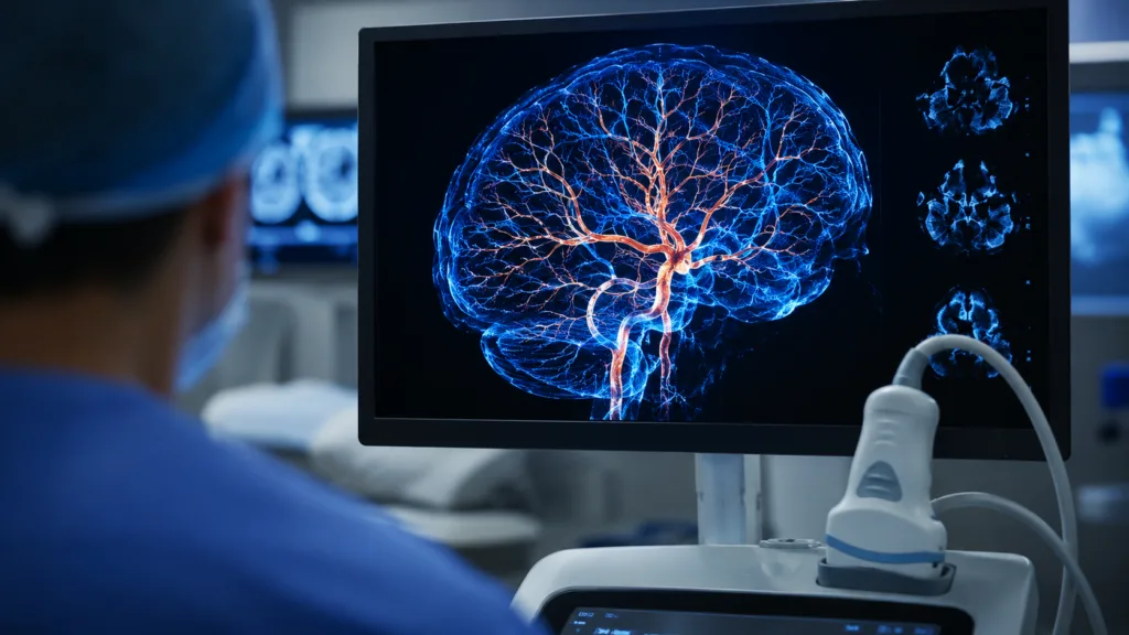
Ultrasound localization microscopy extends that proposition by tracking individual contrast microbubbles rather than relying only on conventional reflected ultrasound. By accumulating millions of bubble positions, the technique can reconstruct vessels smaller than the normal wavelength-limited resolution of the imaging system. The resulting image may reveal vascular networks that standard ultrasound cannot separate clearly.
The trade-off is that super-resolution reconstruction is computationally intensive, dependent on carefully controlled acquisitions and currently reliant on an injected contrast agent. A visually impressive image does not automatically translate into a fast, repeatable or clinically useful examination. The technology will need to prove that it can deliver consistent results across different operators, skull anatomies, patient populations and disease states before comparisons with established clinical modalities become meaningful.
Why the intact skull remains the decisive technical barrier for transcranial ultrasound systems
The human skull weakens, scatters and distorts ultrasound waves. Bone thickness, curvature and internal structure vary between individuals and across different regions of the head, making it difficult to focus energy accurately or reconstruct signals consistently. This is one reason why many high-resolution functional ultrasound experiments have relied on surgical windows, removed bone sections or animal models with more favourable anatomy.
Aleph Neuro’s ability to produce a three-dimensional vascular reconstruction through an intact human skull is therefore the central technical signal in the announcement. It suggests that semiconductor-based ultrasound hardware, specialised acquisition methods and computational correction can recover more usable information than conventional transcranial ultrasound systems. The result also supports the wider view that progress in medical imaging may increasingly depend on software, raw-data access and signal processing rather than hardware power alone.
However, one successful acquisition does not resolve the skull problem at population scale. Temporal bone windows can be poor in some adults, particularly in older patients and individuals with greater bone density. Head movement, probe positioning, acoustic coupling and anatomical variation can all reduce image quality. A system intended for clinical use would need automated calibration, quality controls and clear failure criteria so clinicians know when an image is trustworthy and when a different modality is required.
There are also safety and engineering constraints. The system must deliver enough acoustic energy to obtain a useful signal without exceeding exposure limits or producing unwanted thermal and mechanical effects. Any future wearable or repeated-use configuration would face additional questions around prolonged exposure, sensor stability and consistent alignment. These challenges are manageable research problems, but they remain central to whether transcranial ultrasound can move from a laboratory result into routine neurological imaging.
What the contrast-enhanced protocol reveals about clinical feasibility and regulatory complexity
The disclosed imaging process used sulfur hexafluoride lipid microspheres infused during a four-minute acquisition. The microbubbles create a strong acoustic contrast because they reflect ultrasound differently from surrounding tissue and blood. Tracking their movement enables the system to map cerebral microvasculature with greater detail than a standard ultrasound image.
The use of an approved ultrasound contrast agent may appear to simplify translation, but approval of the agent does not automatically validate a new brain-imaging indication, infusion method or device combination. Existing United States indications for sulfur hexafluoride lipid microspheres cover defined cardiac, liver and paediatric urinary tract imaging uses. A neurovascular protocol would still require evidence covering dose, administration, imaging parameters, safety monitoring, diagnostic performance and the intended clinical population.
Contrast administration also introduces practical limitations. It requires clinical supervision, screening for contraindications and readiness to manage uncommon but potentially serious hypersensitivity or cardiopulmonary reactions. Those requirements may be acceptable for specialised neurological imaging, but they weaken the vision of a low-friction, consumer-like brain interface that can be used repeatedly outside controlled medical settings.
Aleph Neuro has identified contrast-free neurovascular imaging as the longer-term destination. That would remove one barrier to repeated testing and could improve scalability, but it is technically harder because red blood cells generate a much weaker signal than microbubbles. Machine learning may recover information that conventional processing discards, although any algorithm used to reveal faint vascular or functional patterns would need rigorous protection against artefacts, bias and false reconstruction.
How Butterfly Network’s embedded model could expand ultrasound beyond point-of-care diagnostics
Butterfly Network built its commercial identity around handheld point-of-care ultrasound, where a semiconductor-based probe can cover multiple imaging applications traditionally served by separate transducers. Butterfly Embedded takes the same underlying technology and licenses it to partners developing new devices, imaging architectures and specialised applications.
Aleph Neuro’s work shows why that model could matter. A partner can use Butterfly Network’s chip-level imaging capabilities without reproducing the entire semiconductor, transducer and software stack from the beginning. This may shorten development cycles and allow smaller research groups to concentrate on brain-specific arrays, acquisition protocols, reconstruction software and clinical applications.
For Butterfly Network, the strategic opportunity is broader than selling another handheld probe. Embedded partnerships could create licensing revenue, module demand, development payments and exposure to imaging categories that sit outside conventional point-of-care ultrasound. Neuroimaging, tomography, robotics, catheter-based systems and therapeutic ultrasound could all become platform extensions if partners convert research programmes into regulated products.
The risk is that embedded collaborations transfer much of the commercial uncertainty to early-stage partners. A compelling prototype may never become a cleared device, reimbursed procedure or scalable product. Butterfly Network must also support manufacturing quality, data interfaces and long development timelines while protecting its intellectual property. Aleph Neuro’s images strengthen the platform narrative, but they should not be interpreted as evidence that a commercial neuroimaging market has already been secured.
Why open-source data may accelerate validation but cannot replace controlled clinical evidence
Aleph Neuro has released a processing pipeline and a large sample dataset intended to help researchers reproduce the three-dimensional ultrasound localization microscopy workflow. The pipeline covers beamforming, clutter filtering, bubble detection, localisation, track linking and three-dimensional viewing. This transparency is valuable because imaging claims can be difficult to assess when raw data and reconstruction methods remain proprietary.
Independent researchers can use the released materials to test processing choices, identify artefacts and compare alternative algorithms. Open access may also encourage collaboration between ultrasound physicists, neurologists, machine-learning researchers and medical device developers. For an emerging modality, that can be more useful than publishing only selected images or summary performance claims.
Reproducibility of software, however, is not the same as reproducibility of the medical result. Researchers may be able to regenerate an image from the same dataset while remaining unable to acquire comparable data from another participant, scanner or clinical site. True validation will require prospective studies with predefined protocols, multiple participants, independent operators and comparison against recognised reference methods.
Peer-reviewed evidence will also need to describe the study population, acquisition success rate, excluded scans, image-quality thresholds and statistical analysis. Without those details, it is difficult to determine whether the disclosed image represents typical performance or an unusually favourable case. Open sourcing is a constructive first step, but clinical credibility will depend on new data rather than repeated processing of a single dataset.
What must happen before neurovascular ultrasound can compete with MRI, CT or electrophysiology
A new imaging modality does not need to replace every established technology to become commercially valuable. It may succeed by solving a narrower problem more quickly, cheaply or conveniently. Transcranial neurovascular ultrasound could initially find a role in research laboratories, intensive care units, stroke monitoring, bedside assessment or repeated functional studies where conventional scanners are difficult to access.
The strongest development path would begin with a clearly defined clinical question. A system could be evaluated for detecting a vascular abnormality, monitoring a known condition or measuring a change over time. Those tasks offer measurable endpoints and comparison standards. A broad promise to visualise the brain or enable telepathic communication is much harder to validate and risks obscuring the nearer-term medical opportunity.
Clinical studies would need to compare ultrasound findings with magnetic resonance angiography, computed tomography angiography, perfusion imaging or another appropriate reference. Developers would also need to demonstrate repeatability, sensitivity, specificity, acquisition time and performance in patients with difficult acoustic windows. A technically detailed image is useful, but a clinical device must help clinicians make a decision more accurately, more rapidly or at lower total cost.
Commercial adoption would bring another set of requirements. Hospitals would need compatible workflows, trained staff, cybersecurity protections, data storage and integration with imaging archives. Payers would need evidence that the technology improves outcomes or reduces costs. Manufacturers would need reliable production, calibration and service infrastructure. None of these issues diminishes the scientific milestone, but they determine whether the technology becomes a medical product rather than a research achievement.
What clinicians and regulators will watch as Aleph Neuro moves toward contrast-free imaging
The next meaningful evidence will be less about producing an even more striking image and more about demonstrating consistency. Clinicians will want to see results from multiple human participants, including people of different ages and skull characteristics. They will also look for head-to-head comparisons with established imaging and evidence that the technique detects clinically relevant abnormalities rather than only normal vascular anatomy.
Regulators will focus on intended use. A research system for visualising vascular flow presents a different risk profile from a diagnostic device that identifies stroke, predicts neurodegeneration or interprets brain activity. Claims involving automated decision support, functional decoding or long-duration monitoring would add further software, safety and human-factors requirements.
Industry observers are also likely to examine whether contrast-free imaging can approach the resolution achieved with microbubbles. If machine learning becomes central to that effort, developers will need to show that reconstructed features represent genuine biological signals and remain stable across hardware versions, sites and patient groups. Training data quality may become as important as transducer performance.
Aleph Neuro’s result is best understood as evidence that high-resolution transcranial ultrasound is moving into a more credible engineering phase. It does not yet prove that non-invasive brain interfaces are imminent, but it may broaden the range of vascular and functional information that ultrasound systems can capture through an intact skull. For Butterfly Network, the milestone also offers an early test of whether Ultrasound-on-Chip can evolve from a handheld diagnostic platform into a component for entirely new imaging categories.
