MolecuLight Inc. has highlighted a peer-reviewed feasibility study evaluating the MolecuLightDX fluorescence imaging device as an intraoperative adjunct during revision knee arthroplasty for chronic periprosthetic joint infection. The study, published in BMJ Open on April 15, 2026 and promoted by MolecuLight on July 23, prospectively enrolled 10 patients at the Royal Orthopaedic Hospital NHS Foundation Trust in Birmingham, England. It found that fluorescence-directed sampling performed after standard radical debridement recovered bacterial organisms in eight patients, with the isolates matching those obtained through routine sampling.
The findings provide an early indication that real-time bacterial fluorescence imaging may help surgeons identify tissue that still carries bacterial burden after conventional debridement appears complete. They do not, however, demonstrate that using the device reduces recurrent infection, prevents further revision surgery, improves implant survival or produces better patient outcomes.
That distinction will determine how the study should be interpreted. MolecuLightDX has an established commercial and regulatory position in wound imaging, but its use inside an infected prosthetic knee represents a new clinical application requiring substantially stronger evidence before it can influence routine orthopaedic practice.
What did the MolecuLightDX knee revision feasibility study actually demonstrate?
Researchers recruited 10 patients with established chronic periprosthetic knee joint infection between January and December 2023. The patients ranged from 55 to 79 years old, with a median age of 65.5 years. Six were men and four were women, and all cases were managed through a specialist multidisciplinary periprosthetic joint infection service.
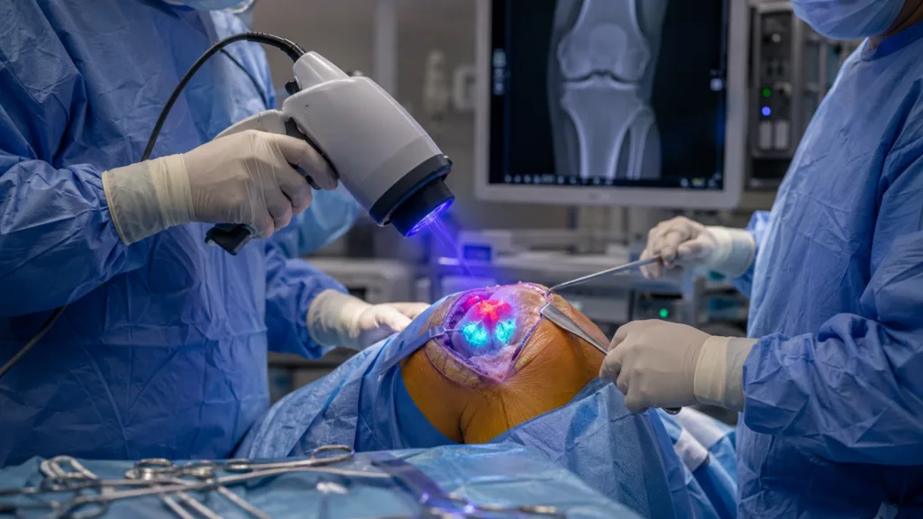
Each patient first underwent the hospital’s standard radical debridement procedure. The surgeon removed tissue considered infected, contaminated or devitalised and collected routine microbiology and histopathology samples. Only after the conventional debridement was considered complete was MolecuLightDX used to inspect the operative field for additional fluorescence.
Fluorescent areas identified by the device were excised using fresh instruments and sent for separate microbiological or histological assessment. The device detected at least one additional fluorescent area in all 10 procedures.
Cultures from the fluorescence-directed samples isolated an organism in eight of the 10 patients. In each of those eight cases, the organism matched the pathogen recovered through standard intraoperative sampling, which reduced the likelihood that the fluorescence-directed results were merely caused by sample contamination.
Histological samples were obtained from fluorescent tissue in eight cases. Three showed findings meeting the study’s definition of acute infection, while five showed inflammatory tissue considered consistent with chronic infection. The study therefore produced microbiological and histological evidence that tissue selected through fluorescence could contain residual infection-related material even after experienced surgeons had completed standard debridement.
The study did not include statistical hypothesis testing because it was designed only to determine feasibility. The results should consequently be read as an early signal supporting further investigation, not as a quantified estimate of diagnostic accuracy or clinical effectiveness.
Why does detecting matching bacterial isolates matter without proving better surgical outcomes?
Periprosthetic joint infection is among the most difficult complications associated with joint replacement. Successful treatment depends on several interconnected steps, including identifying the infecting organism, selecting appropriate antimicrobial therapy, removing infected tissue and determining whether implants can be retained or must be exchanged.
The adequacy of surgical debridement is particularly difficult to measure in real time. Surgeons rely on visual assessment, tissue appearance, anatomical knowledge and clinical experience, but bacterial burden cannot normally be seen with the naked eye. This creates the possibility that apparently acceptable tissue may still contain organisms capable of contributing to persistent or recurrent infection.
The MolecuLight study addresses this practical problem by testing whether fluorescence could reveal additional areas for sampling and removal. Recovering the same organisms from both routine samples and fluorescence-directed samples suggests that the signals were associated with the known infection rather than unrelated environmental contamination.
However, microbiological concordance is not equivalent to clinical utility. The study did not compare fluorescence-guided surgery with standard surgery in a control group. It also did not establish whether the additional tissue removal was necessary, whether it changed antimicrobial treatment or whether patients receiving fluorescence-guided debridement experienced fewer recurrences.
Some fluorescent tissue could contain bacteria that would already have been controlled by antibiotics or the broader surgical procedure. Conversely, a lack of fluorescence cannot be assumed to exclude residual infection. The device is therefore better understood as a potential adjunct to surgical judgement rather than a definitive method of identifying every infected area.
How does MolecuLight fluorescence imaging identify bacterial signals without an injected dye?
MolecuLight’s platform uses violet excitation light at a wavelength of approximately 405 nanometres to stimulate naturally occurring fluorescent compounds in tissue and bacteria. Unlike fluorescence systems that require an injected or topically applied contrast agent, MolecuLight relies on endogenous autofluorescence.
Porphyrins produced by several bacterial species can generate red fluorescence, while pyoverdines associated with Pseudomonas aeruginosa can produce cyan fluorescence. Structural tissue components such as collagen and fibrin generally produce green fluorescence, providing anatomical context for interpreting the image.
In its established wound-care application, the device is intended to help clinicians identify and document areas associated with elevated bacterial loads. Fluorescence is interpreted alongside clinical signs, symptoms and professional judgement. It does not independently diagnose infection, determine antibiotic susceptibility or replace microbiological culture.
The technology has practical characteristics that could appeal to orthopaedic teams. It is handheld, provides information during the procedure and does not require surgeons to wait for laboratory results before deciding whether to remove additional tissue. Avoiding an injected imaging agent could also simplify the procedure compared with contrast-dependent fluorescence techniques.
Operating inside a prosthetic joint nevertheless presents a different environment from imaging an exposed skin wound. Blood, bone, scar tissue, implant surfaces, tissue depth, ambient lighting and inflammatory changes could all affect signal visibility or interpretation. Protocols developed for chronic wound imaging cannot automatically be assumed to perform identically in revision arthroplasty.
What are the most important limitations of the 10-patient orthopaedic study?
The most obvious limitation is scale. Ten patients from a single specialist centre cannot represent the wide range of organisms, implants, host factors, previous surgeries and infection patterns encountered across orthopaedic practice.
All debridement procedures were performed by one experienced periprosthetic joint infection surgeon. This provided procedural consistency but limits generalisability. Results could differ when the technology is used by surgeons with varying experience, different debridement techniques or less familiarity with fluorescence interpretation.
The study was also unblinded. The operating surgeon could see the fluorescence and made subjective decisions about which signals warranted additional sampling. Fluorescence interpretation was not standardised, meaning the study did not establish an objective threshold that could be reproduced reliably across hospitals and operators.
There was no control group, randomisation or independent blinded assessment of images. The research consequently cannot determine whether fluorescence-guided debridement performs better than conventional surgery or whether another experienced surgeon would have removed the same tissue without imaging.
Most importantly, the study lacked clinical outcome data. It was not designed to measure infection eradication, reinfection, reoperation, functional recovery, implant retention, hospital length of stay or the duration of antimicrobial treatment. The authors acknowledged that the small sample and methodological design prevented statistical analysis or broader interpretation.
MolecuLight supplied the device and supported the article’s publication charges, although the study was conducted by Royal Orthopaedic Hospital researchers and the authors declared no competing interests. That disclosure does not invalidate the findings, but independent replication without manufacturer support would strengthen confidence in the technology.
Why does MolecuLightDX’s current regulatory status require careful interpretation?
MolecuLightDX is a United States Food and Drug Administration-cleared Class II device. The agency classifies it as an autofluorescence imaging adjunct tool for wounds under 510(k) number K211901, with its regulatory specialty listed under general and plastic surgery.
Its stated United States intended use covers clinicians diagnosing and treating skin wounds at the point of care. The device can capture wound images, measure wound dimensions and record fluorescence emitted from wounds exposed to excitation light.
The fluorescence image, when combined with clinical signs and symptoms, is intended to increase the likelihood of identifying wounds containing bacterial loads above the specified threshold. The manufacturer’s intended-use information explicitly states that the technology should not be used to rule out bacteria and does not itself diagnose or treat skin wounds.
The BMJ Open study does not constitute regulatory clearance for intraoperative use during revision knee arthroplasty. It also does not represent a new orthopaedic indication, a label expansion or confirmation that the device is clinically effective in prosthetic joint infection surgery.
MolecuLight would need to determine the appropriate regulatory strategy for any formal expansion into orthopaedic applications. Regulators would likely examine device performance in the surgical environment, interpretation consistency, false-positive and false-negative risks, usability, sterility controls and whether fluorescence-guided decisions improve clinically meaningful outcomes.
What would a larger trial need to prove before fluorescence-guided debridement enters routine practice?
The next evidence step should move beyond demonstrating that fluorescence can be observed. A multicentre prospective study would need to establish whether different surgeons can interpret the images consistently and whether fluorescence accurately identifies tissue containing clinically relevant bacterial burden.
A controlled design comparing standard debridement with fluorescence-guided debridement would be particularly valuable. Relevant endpoints could include infection eradication, repeat surgery, implant retention, antibiotic duration, complications, functional recovery and healthcare utilisation.
Researchers would also need to define how fluorescence findings should change surgical behaviour. Removing every fluorescent area without a validated protocol could lead to unnecessary excision of viable tissue, potentially increasing operative complexity or compromising reconstruction. Ignoring ambiguous signals could create the opposite problem.
Training will therefore be central. Hospitals would need standard procedures covering device preparation, lighting, sterility, image acquisition, signal interpretation, documentation and the relationship between fluorescence, culture and histology.
Health-economic evidence would also matter. Avoiding even a small number of recurrent infections or repeat revisions could create substantial value, but that benefit cannot be assumed from a feasibility study. Procurement teams will expect evidence that equipment, training and additional operating-room time are justified by measurable improvements in outcomes or costs.
Could orthopaedic surgery become a meaningful commercial expansion for MolecuLight?
MolecuLight is a privately held medical imaging company that currently manufactures and commercialises the MolecuLight i:X and MolecuLightDX platforms primarily for wound assessment. The company has built its evidence base around bacterial fluorescence imaging, wound measurement and point-of-care documentation.
A validated orthopaedic application could broaden the platform beyond outpatient wound clinics and specialist wound services into operating rooms, revision centres and bone-infection programmes. The commercial importance would depend on whether the existing hardware can be adapted to surgical workflows without extensive redesign and whether the company can secure an appropriately defined regulatory indication.
The 10-patient study provides MolecuLight with a scientific starting point rather than a near-term commercial endpoint. It supports the biological and operational rationale for testing the device in surgery, but it does not yet establish the reproducibility, clinical utility or economic value required for broad hospital adoption.
The decisive milestone will be a larger controlled investigation showing that fluorescence-guided debridement changes more than sampling behaviour. MolecuLight must demonstrate that identifying and removing additional fluorescent tissue produces a meaningful improvement in infection control without causing unnecessary tissue loss or adding disproportionate complexity to revision surgery.
Until such evidence emerges, the BMJ Open publication should be viewed as a credible feasibility signal in a difficult surgical problem. It has opened a new research pathway for MolecuLightDX, but the clinical and commercial case will depend on whether real-time fluorescence can ultimately reduce the recurrence and treatment burden of periprosthetic joint infection.