
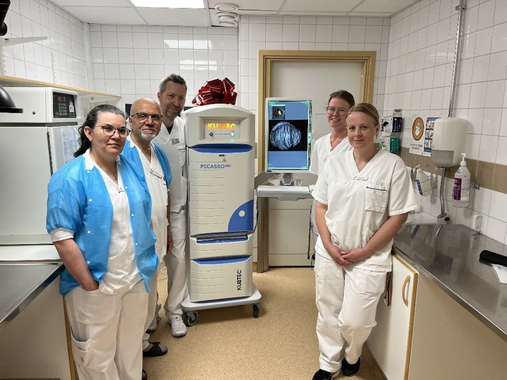
KUB Technologies, Inc., which operates as KUBTEC, has installed its Picasso Plus specimen radiography system at Falu Hospital in Sweden, describing the deployment as the first European installation of the platform. The system has been placed in the hospital’s mammography X-ray department, where radiologists will initially review specimen images and pathologists will be able to access them when required.
The July 28, 2026 announcement marks an important commercial step for a system launched in the United States in February 2025. However, the value of the installation will ultimately be measured less by its “first in Europe” status and more by whether it improves coordination between mammography, radiology and pathology teams handling breast biopsies and surgical specimens.
That distinction matters because KUBTEC has primarily positioned Picasso Plus as a pathology-laboratory platform designed to bring X-ray imaging closer to the grossing workflow. At Falu Hospital, the equipment is operating within mammography, creating a different implementation model in which radiologists remain the first readers while pathology personnel gain access to the images. The arrangement could provide a useful test of how the platform functions across departmental boundaries rather than exclusively inside a pathology gross room.
Why does the Falu Hospital installation matter beyond being KUBTEC’s first Picasso Plus site in Europe?
A single installation does not establish broad European adoption, but the selection of Falu Hospital gives KUBTEC a potentially valuable reference site. Falu Hospital is the largest hospital in Dalarna County and serves as the centre for specialist healthcare in the region. Region Dalarna says the hospital has approximately 450 beds and around 3,300 staff members providing medical and surgical care.
The hospital’s pathology and cytology operation also serves the wider county, covering histopathology, cytology and specialised diagnostic procedures. This regional role means that improvements in specimen handling or radiology-pathology coordination may have implications beyond an individual department, particularly when complex breast cases require correlation between imaging findings, gross examination and microscopic diagnosis.
Falu Hospital already has established clinical mammography services and experience with multimodal breast cancer assessment. Region Dalarna’s published information describes mammography in Falun as part of its diagnostic imaging organisation, while local clinical research has examined the relationship between breast imaging, tumour distribution and large-section histology. The environment therefore appears suited to evaluating a platform intended to connect radiographic findings with subsequent pathological assessment.
KUBTEC said clinical staff found Picasso Plus easy to operate during the first cases and reported that the system displayed microcalcifications even in small biopsies held in formalin-filled plastic containers. Those observations are encouraging as early user feedback, although they remain site-level comments rather than results from a prospective workflow or diagnostic-performance study.
How will the mammography-led workflow shape the system’s practical value at Falu Hospital?
KUBTEC markets Picasso Plus as a system capable of reducing the need to transport specimens from pathology to radiology or mammography. That potential benefit is most direct when a system is physically installed in the gross room and operated by pathology personnel during specimen preparation. At Falu Hospital, the system’s location in mammography means the workflow may retain at least some movement of specimens or coordination between departments.
This does not necessarily diminish the value of the installation. Radiologist-first review may fit the hospital’s existing governance, staffing and image-interpretation practices, particularly where radiographic confirmation of microcalcifications or surgical markers already sits within mammography. It may also allow the hospital to introduce the technology without immediately redesigning the pathology laboratory or transferring image-review responsibilities.
The trade-off is that some of the efficiencies promoted for gross-room deployment will need to be demonstrated within the hospital’s actual configuration. Procurement teams and clinical managers are likely to examine how quickly specimens move through the imaging process, whether images are readily available to pathology staff, whether repeat imaging declines and whether the system reduces delays in selecting tissue for histological processing.
The deployment also highlights an important reality for medical device commercialisation. Hospitals do not always implement technology exactly as manufacturers envision it. Local staffing models, radiation-safety arrangements, room availability, information technology infrastructure and established clinical responsibilities can produce hybrid workflows. A successful European expansion strategy may therefore require KUBTEC to support several operating models rather than promoting a single standard installation.
Why is specimen radiography important when breast biopsies contain clips and microcalcifications?
Specimen radiography is commonly used to confirm that tissue removed during a breast biopsy or surgical procedure contains the imaging target that prompted the intervention. These targets can include microcalcifications, localisation markers and surgical clips that may not be readily visible during gross examination.
The clinical importance lies in radiological-pathological correlation. When an abnormality identified on mammography leads to biopsy, teams must confirm that the sampled tissue contains the expected target and that the histological findings adequately explain the imaging appearance. A mismatch can require additional tissue examination, deeper sectioning, further radiographic review or, in some circumstances, another procedure.
Published research has long supported the use of specimen radiography in biopsies performed for mammographically detected microcalcifications. One Radiology study involving 361 stereotactic breast core specimens found that histological detection of calcifications was substantially more frequent when calcifications were visible on the specimen radiograph. The authors concluded that specimen radiography helped confirm appropriate sampling and direct pathological examination.
More recent reviews continue to emphasise the need to radiograph fragments obtained from biopsies performed for microcalcifications and to correlate the radiographic findings with the samples selected for pathology. They also underline that calcifications can be associated with benign conditions, lesions of uncertain potential, ductal carcinoma in situ and invasive malignancy, making careful correlation more important than image resolution alone.
That final point is essential when assessing the commercial claims surrounding advanced specimen imaging. Producing a sharper image or identifying more calcifications does not independently establish better diagnostic accuracy, fewer repeat procedures or improved patient outcomes. One microradiography study found that substantially higher image resolution did not produce a statistically significant improvement in diagnostic validity over conventional specimen radiography.
Picasso Plus should therefore be understood as a workflow and visualisation tool supporting professional interpretation, specimen localisation and tissue selection. It does not replace histopathology, multidisciplinary review or the clinical judgement required to determine whether radiological and pathological findings are concordant.
What differentiates Picasso Plus, and where does the supporting evidence remain incomplete?
KUBTEC says Picasso Plus uses amorphous selenium direct-capture technology, an approach also used in digital mammography detectors. The company has configured the system with a 10-inch by 12-inch imaging area, five magnification levels and an optical camera that produces a visual image of the specimen alongside the X-ray image.
Its Image Blender feature combines the optical and X-ray images, potentially helping users relate radiographic findings to the specimen’s visible orientation. The intended workflow includes locating clips, margins and microcalcifications before tissue is selected for placement into histology cassettes. The system also offers voice control, an optional portable tablet, image-export functionality and an alert intended to notify users when a specimen has been left inside the cabinet.
These features address practical problems encountered during specimen handling. Optical and radiographic image alignment may make it easier to communicate the location of a target, while direct image export could reduce manual steps if it integrates reliably with the hospital’s laboratory information system.
However, the published announcement did not disclose controlled clinical data comparing Picasso Plus with the system previously used at Falu Hospital. It did not provide measurements for turnaround time, repeat imaging, tissue-block selection, reader agreement, diagnostic concordance or the number of cases requiring additional processing.
The announcement also did not specify the European conformity assessment or CE-marking documentation supporting the installation. Medical device manufacturers are responsible for ensuring that devices placed on the European market meet the applicable conformity and CE-marking requirements under the European regulatory framework. The absence of these details from the announcement should not be interpreted as evidence of a regulatory deficiency, but publishing the relevant status would make the device’s European commercial position clearer.
Can one Swedish installation help KUBTEC build a wider European pathology imaging business?
KUBTEC is a family-owned medical imaging company with an established portfolio of specimen radiography systems for operating rooms, biopsy suites and pathology laboratories. Picasso Plus broadens that portfolio by targeting pathology workflows that have historically depended on imaging equipment located elsewhere in a hospital.
For a specialised capital-equipment manufacturer, the first regional installation can have commercial significance beyond its immediate revenue. Prospective customers often want to observe a comparable hospital using the system, speak with clinical users and understand installation, training, servicing and integration requirements before beginning their own procurement process.
Falu Hospital could provide that reference, particularly for Nordic healthcare systems with centralised regional services and formal multidisciplinary breast cancer pathways. Nevertheless, converting a first installation into a repeatable European business will require more than favourable staff feedback.
KUBTEC will need reliable local servicing, application training and technical support. Hospitals will also evaluate acquisition cost, maintenance arrangements, detector lifespan, software support, cybersecurity, information-system compatibility and the volume of cases that justify dedicated specimen imaging.
The location of the Falun system may become especially relevant to future sales discussions. Some hospitals may prefer the pathology-centred workflow promoted by KUBTEC, while others may retain radiologist-led review within mammography. Demonstrating that Picasso Plus can work effectively under both models would give the company greater flexibility when approaching European institutions.
What will determine whether Picasso Plus becomes a scalable European hospital platform?
The next meaningful evidence will come from the system’s performance in routine practice. Useful measures would include image acquisition time, specimen transport time, the frequency of repeat radiographs, the speed of locating clips or calcifications and the proportion of cases in which imaging changes tissue-block selection.
Hospitals will also want to know whether access to optical and radiographic images improves communication between radiologists, pathologists and pathology assistants. Any reduction in turnaround time must be interpreted alongside staffing requirements and the possibility that adding another imaging step could create a new bottleneck if responsibilities are not clearly defined.
For KUBTEC, the Falun installation is best viewed as a commercial entry point and implementation test rather than confirmation of widespread European demand. The company has placed a relatively new platform inside a large regional hospital with established mammography and pathology capabilities, giving it an opportunity to show how the technology performs in a clinically sophisticated setting.
The decisive question is now whether the system can generate measurable workflow benefits under Falun’s radiologist-first configuration. Evidence from that experience, combined with transparent regulatory information, reliable service support and additional European installations, will determine whether Picasso Plus develops from a notable first deployment into a repeatable pathology imaging business.