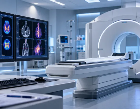
Cancer treatment has traditionally divided imaging and therapy into separate activities: one technology locates the disease and another attempts to destroy it. Radiotheranostics collapses much of that distinction by using related molecular compounds to first image a biological target and then deliver therapeutic radiation to cells carrying the same target. Instead of irradiating an anatomical area from outside the body, a radiopharmaceutical can travel systemically, bind a molecular marker on cancer cells and carry a radioactive isotope directly to metastatic sites distributed throughout the patient.
The strategy has already moved from specialist nuclear-medicine practice into mainstream oncology through lutetium-177 therapies targeting somatostatin receptors in neuroendocrine tumors and prostate-specific membrane antigen in prostate cancer. Development is now spreading toward alpha emitters such as actinium-225 and lead-212, alternative beta emitters such as terbium-161 and entirely new molecular targets. The scientific opportunity is large, but radiopharmaceuticals also introduce challenges conventional pills and monoclonal antibodies rarely face: isotopes decay continuously, manufacturing and administration are time-sensitive, specialized facilities are required and the supply of certain radionuclides can become part of the medicine’s clinical availability.
How can the same biological target be used for both diagnosis and treatment?
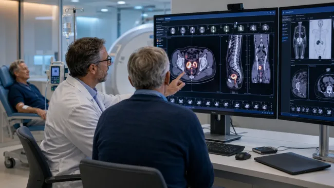
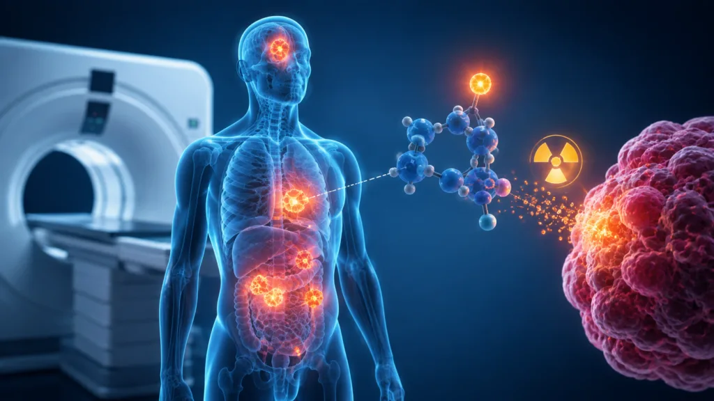
A theranostic strategy usually begins with a targeting molecule designed to recognize a protein or receptor enriched on cancer cells. For diagnostic imaging, that molecule is coupled to a radionuclide whose emissions can be detected using PET or another nuclear-imaging system. The resulting scan shows whether tumors across the patient’s body express enough of the target to justify therapy and can simultaneously reveal sites of disease that might not be obvious from anatomy alone.
The therapeutic version uses the same or a closely related targeting vector attached to an isotope capable of delivering cell-damaging radiation. A well-established example is PSMA-targeted prostate cancer treatment, where patients undergo PSMA imaging to determine whether their tumors express the target before receiving a PSMA-directed lutetium-177 therapy. FDA has continued expanding the clinical reach of this model, including a 2026 approval of lutetium Lu 177 vipivotide tetraxetan with androgen-receptor pathway therapy in an earlier PSMA-positive metastatic prostate cancer population.
This creates an unusual form of personalized medicine because clinicians can visualize the therapeutic target across the entire body before committing to treatment. A biopsy samples only one location and can miss heterogeneity between metastases, whereas whole-body molecular imaging can reveal whether target expression varies dramatically between lesions.
Why has lutetium-177 become the current benchmark isotope?
Lutetium-177 emits beta particles capable of traveling through tissue beyond the individual cell carrying the radiopharmaceutical. This produces a crossfire effect in which radiation can damage nearby tumor cells even if uptake is not perfectly uniform. That property is useful in larger or heterogeneous lesions because every malignant cell does not necessarily have to bind the same amount of radioligand for surrounding tissue to receive therapeutic radiation.
The isotope also supports post-treatment imaging, helping nuclear-medicine teams visualize where therapy has distributed. Combined with established production and clinical experience, these characteristics have helped lutetium become the leading radionuclide in modern targeted radiopharmaceutical therapy. Randomized evidence in prostate and neuroendocrine cancers has created a regulatory and operational foundation on which newer programmes can build.
Beta particles are not ideal for every biological problem, however. Their relatively longer tissue range distributes radiation over multiple cell diameters, which can be advantageous for bulky disease but may be less efficient against tiny clusters or individual malignant cells. Resistance to a lutetium-labelled therapy can also occur because of changing target expression and other tumor characteristics.
Why are alpha emitters such as actinium-225 attracting so much attention?
Alpha particles travel far shorter distances than beta particles but deposit considerably more energy over that path. The result is extremely dense DNA damage concentrated within a few cell diameters, creating a strong theoretical advantage for microscopic disease and cancer cells that remain target-positive despite resistance to beta-emitting therapy. Actinium-225 has therefore become one of the most closely watched radionuclides in PSMA-directed development.
The trade-off is that high-energy alpha radiation can also cause severe toxicity when radiopharmaceuticals accumulate in healthy organs. Salivary-gland toxicity has been a particular concern in PSMA-targeted actinium programmes, while dosimetry and imaging of alpha emitters can be more complicated than for lutetium. Prospective controlled evidence is also far less mature, meaning impressive early responses should not be treated as proof that alpha therapy is broadly superior.
Supply creates another constraint. Actinium-225 is difficult to manufacture at the scale that could eventually be required for global oncology use, turning isotope-production investment into part of the competitive strategy around the entire drug class. Other radionuclides including lead-212 and terbium-161 are being studied partly because their physical properties and production pathways could address different clinical and logistical limitations.
Why is manufacturing a radiopharmaceutical different from manufacturing a conventional cancer drug?
A bottle of tablets can be manufactured, stored and shipped months before a patient needs it. Radioactive isotopes are continuously decaying from the moment they are produced, meaning the product is effectively losing activity while it moves through the supply chain. Manufacturing therefore has to coordinate isotope production, radiolabelling, quality release, transportation and the patient’s appointment within a tightly controlled window.
This creates unusual infrastructure requirements. Radiopharmaceutical production needs radiation-handling capabilities, specialized quality systems and reliable access to radionuclides, while hospitals require appropriately trained nuclear-medicine teams, radiation-safety procedures and facilities capable of administering and managing radioactive treatments. Recent expert reviews have emphasized that expansion of theranostics depends not only on successful drug trials but also on workforce and operational readiness.
The supply chain can therefore become a competitive advantage. A scientifically excellent radioligand is commercially limited if a company cannot secure enough isotope, label it reliably and deliver patient-specific doses to treatment centers before radioactive decay makes them unusable.
Could dosimetry eventually make radiopharmaceutical treatment more personalized?
External-beam radiation oncology routinely calculates how much radiation is delivered to a tumor and surrounding organs. Radiopharmaceutical therapy has historically relied more heavily on standardized administered activities, but imaging before and after treatment can potentially allow physicians to estimate how much radiation particular organs and tumors actually receive.
This creates an opportunity for patient-specific dosimetry. Two people receiving the same injected activity can distribute a radiopharmaceutical differently because of tumor burden, renal clearance, target expression and normal-organ uptake. Measuring that distribution may eventually allow treatment to be intensified in patients whose healthy tissues can tolerate more radiation or reduced when normal organs are receiving excessive exposure.
The practical challenge is complexity. Repeated imaging, quantitative reconstruction, specialized software and medical-physics expertise add work to treatment pathways that are already resource-intensive. Whether personalized dosimetry becomes routine will depend on evidence that it improves outcomes enough to justify the added operational burden.
Will radiopharmaceutical therapy replace chemotherapy or external radiation?
It is more likely to become another major treatment modality than to eliminate existing ones. Radiopharmaceutical therapy is uniquely suited to disseminated cancers carrying an accessible molecular target, but tumors can lose that target, express it unevenly or contain disease components that do not accumulate enough radioligand for adequate radiation delivery.
Combination therapy is therefore likely to become increasingly important. Radiopharmaceuticals can potentially be combined with hormonal therapy, DNA-damage repair inhibitors, immune therapies or conventional radiation depending on disease biology, while external-beam treatment retains advantages for precise control of defined anatomical targets.
The most durable idea behind theranostics is not any single isotope. It is the ability to ask a molecular question before treatment: does this patient’s cancer express enough of the target for the therapeutic drug to reach it? The diagnostic scan provides the answer, while the therapeutic radiopharmaceutical uses the same biology to deliver radiation. That combination makes radiotheranostics one of the clearest examples of precision medicine moving beyond selecting a drug from a laboratory test and into directly visualizing where a treatment is likely to travel inside an individual patient.