Hyperfine Inc. has received another US Food and Drug Administration clearance for its Optive AI software, adding improved noise correction, a new isotropic T1 sequence and workflow changes to the Swoop portable magnetic resonance imaging system as the company attempts to narrow the practical image-quality gap between bedside ultra-low-field MRI and conventional scanners installed in radiology departments. Hyperfine plans to begin deploying the latest software to new and existing US Swoop customers in September 2026.
The update is Hyperfine’s second FDA-cleared software package since it launched the redesigned Swoop system in mid-2025 and continues a rapid regulatory cadence around the platform. Earlier iterations introduced the Optive AI reconstruction architecture and subsequently a multidirectional diffusion-weighted imaging sequence intended to strengthen stroke visualization. FDA records identify Swoop as a Class II nuclear magnetic resonance imaging system, and earlier cleared versions have entered through the 510(k) substantial-equivalence pathway.
The strategic question is increasingly less about whether a portable MRI scanner can produce clinically useful brain images and more about how many situations can be shifted away from the conventional MRI suite. Swoop operates at ultra-low magnetic-field strength and can be wheeled to professional-care settings such as intensive care units and emergency departments, allowing selected patients to be imaged without transportation to a fixed scanner.
Hyperfine’s latest software is therefore important because image quality remains the central trade-off behind portable low-field imaging. Mobility, lower infrastructure requirements and bedside access are valuable only if clinicians receive enough anatomical information to answer the clinical question that prompted imaging.
What does the new Optive AI software change compared with earlier Swoop versions?
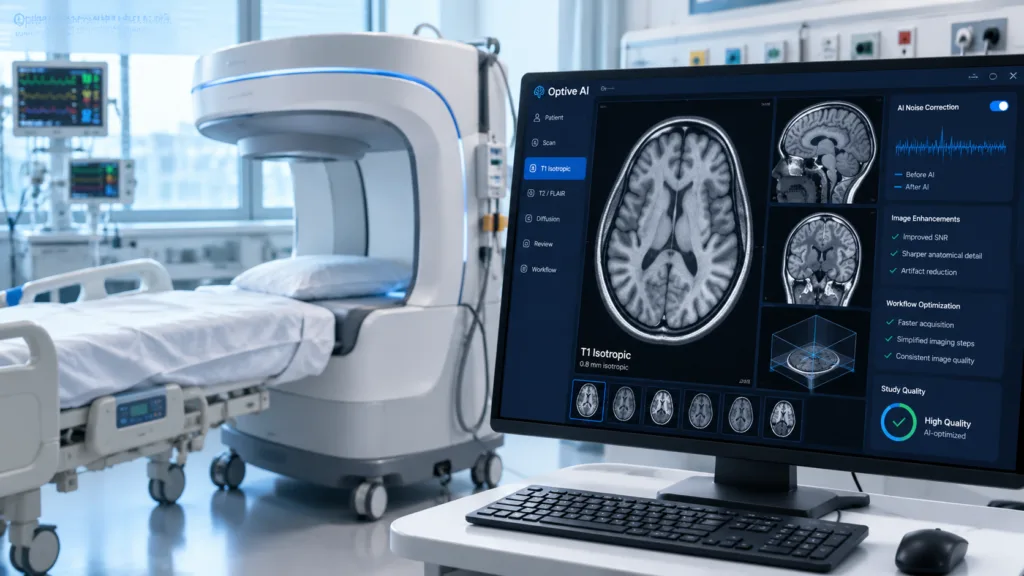
The update expands Hyperfine’s proprietary noise-cancellation technology across imaging sequences, targeting electromagnetic interference that can become particularly relevant when MRI is used outside heavily engineered radiology environments. Unlike conventional high-field scanners that depend on dedicated shielded rooms and tightly controlled siting, Swoop is designed to function in environments such as intensive care units and emergency departments where electrical equipment and other sources of interference are common.
Hyperfine has also redesigned parts of the operator interface. The software provides a more guided scanning workflow and allows sequences and images to be viewed while acquisition is still underway, potentially helping operators recognize problems before the entire examination is completed.
The more technically interesting change is the isotropic T1 sequence. Isotropic imaging uses voxels with equivalent dimensions across three spatial axes, allowing datasets to be reconstructed and examined in different planes without the same loss of detail associated with thicker direction-dependent slices.
Hyperfine said the new sequence is intended to provide thinner slices and comparable resolution across imaging planes, creating a foundation for three-dimensional visualization. The company highlighted postoperative tumor assessment as one potential setting where spatial representation could be useful, while describing contrast-enhanced neurosurgery applications and quantitative MRI as possible future directions rather than currently established indications.
That distinction is important. FDA clearance of the software update does not mean Swoop has received authorization for every future application Hyperfine is investigating. The system remains cleared for brain imaging where a full diagnostic examination is not clinically practical, and images are intended to be interpreted by a trained physician.
How much evidence has accumulated for portable MRI outside a conventional radiology department?
Hyperfine’s commercial argument has increasingly been supported by clinical studies examining workflow rather than only technical image quality. The prospective NEURO PMR programme compared portable MRI with standard-of-care high-field imaging in outpatient neurology practices, examining whether Swoop-generated information was clinically useful across common neurological presentations and how patients experienced the examination.
The company has separately examined emergency-department use through the PRIME study. Hyperfine reported in May that the prospective randomized study evaluated bedside portable MRI in patients presenting with neurological emergencies, with a focus on time to imaging and usefulness for triage. Those results were presented at the Society for Academic Emergency Medicine meeting and should be distinguished from completed regulatory evidence for a new diagnostic indication.
Stroke represents another important use case because rapid brain imaging can affect clinical decisions while moving critically ill patients through a hospital introduces logistical delays. A multicenter observational dataset involving 95 patients assessed Swoop stroke detection using participants from Massachusetts General Hospital, Buffalo General Medical Center and Yale New Haven Hospital. Hyperfine reported the work as the largest Swoop stroke dataset available at that point.
The company has also accumulated evidence in neurosurgical environments. Peer-reviewed publications highlighted in July examined use of portable MRI after endovascular neurosurgical procedures, including imaging performed in ambulatory settings before same-day discharge. Those reports demonstrate potential workflow applications but do not establish that ultra-low-field MRI should replace conventional MRI or CT across all postoperative patients.
Why is noise correction so important for an MRI scanner designed to move around a hospital?
Conventional MRI facilities are built around magnetic-field control. Shielded rooms, restricted equipment and carefully managed infrastructure help reduce environmental interference and protect image quality. Portable MRI reverses part of that model by taking the scanner into clinical environments that were never designed around an MRI magnet.
That flexibility creates both the opportunity and the engineering challenge. Intensive care rooms contain monitors, infusion pumps, ventilators and other electrical systems, while emergency departments are dynamic spaces where perfectly controlled electromagnetic conditions are unrealistic.
Software therefore becomes unusually important. An ultra-low-field device cannot simply reproduce the hardware strategy of a high-field scanner at a fraction of the field strength and expect identical output. Reconstruction algorithms, denoising, acquisition strategy and correction for environmental interference become part of the core imaging architecture.
Hyperfine has been using artificial intelligence partly to compensate for those physical limitations. Its initial Optive AI release applied algorithms across noise cancellation, acquisition, reconstruction and post-processing, while the latest generation extends noise correction and adds a more sophisticated three-dimensional acquisition option.
The relevant comparison nevertheless remains clinical adequacy, not whether an ultra-low-field image looks identical to a 1.5-tesla or 3-tesla scan. Conventional high-field MRI retains major advantages in signal, resolution and advanced sequences. Swoop’s proposition is that sufficiently useful information obtained immediately at the bedside can sometimes be more valuable than technically superior imaging that requires transportation, scheduling and dedicated infrastructure.
Could software upgrades materially change the economics of Hyperfine’s installed Swoop fleet?
Frequent software clearance can create an important commercial effect for a connected imaging platform because improvements can potentially be distributed across existing scanners rather than requiring customers to purchase entirely new capital equipment each time image reconstruction advances.
Hyperfine demonstrated that strategy with earlier Optive AI releases, rolling upgraded software across parts of the installed base after FDA clearance. The new update is likewise planned for both existing and new customers beginning in September.
That model can improve the value proposition for hospitals considering a system whose underlying magnetic hardware is less powerful than conventional MRI. If image quality and clinical applications improve through software, the useful capability of the installed scanner can expand after purchase.
However, regulatory clearance remains necessary when software modifications materially affect the device. Artificial intelligence does not turn an MRI scanner into an unrestricted software platform where every new sequence or diagnostic claim can simply be downloaded commercially without regulatory assessment.
Hyperfine’s history illustrates this clearly. FDA clearance of the original portable system has been followed by multiple submissions covering subsequent generations, Optive AI reconstruction, redesigned hardware and advanced diffusion imaging. The latest clearance extends that pattern rather than replacing it.
Where could portable MRI expand next?
Hyperfine is investigating several directions that would make portable MRI more useful beyond its current role. Contrast-enhanced imaging could materially expand neurological and neurosurgical applications, and the company has been conducting its Contrast PMR study to support a future FDA 510(k) submission involving gadolinium-based contrast agents. That work remains developmental and should not be confused with an already cleared contrast indication.
International expansion is happening alongside US development. The next-generation Swoop system has received European regulatory approvals and entered commercial rollout in Europe, while All India Institute of Medical Sciences, New Delhi began clinical use of the portable scanner in India during 2026.
The latest Optive AI clearance therefore looks less like an isolated software update and more like another incremental attempt to expand the number of clinical questions an ultra-low-field scanner can answer.
The limitation is equally clear: portable MRI does not need to replace high-field MRI to become commercially meaningful, but it does need to prove that bedside access changes patient management often enough to justify the system and its workflow.
Improved noise correction and isotropic T1 imaging directly address that challenge. Each software generation that increases usable anatomical detail without sacrificing mobility makes Swoop more capable, but adoption will ultimately depend on whether hospitals and neurologists find that those incremental gains translate into faster decisions, fewer patient transfers and enough diagnostic confidence to change where brain imaging occurs.