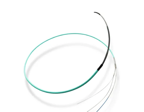
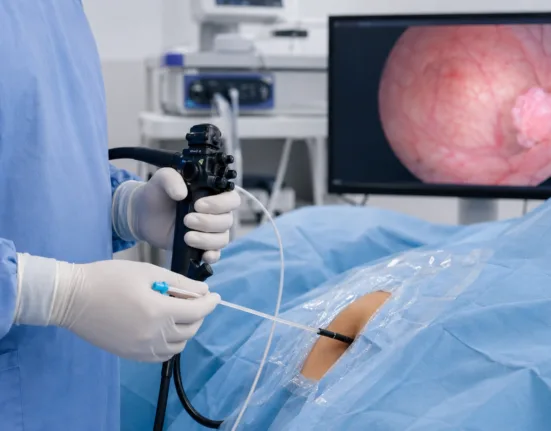
Conavi Medical Corp. has announced a peer-reviewed case publication highlighting the use of its hybrid intravascular ultrasound and optical coherence tomography imaging system in a complex coronary intervention involving a patient with impaired renal function. The publication follows U.S. Food and Drug Administration 510(k) clearance for the Canadian medical device developer’s next-generation hybrid IVUS-OCT imaging system, strengthening the clinical evidence base behind a commercial rollout in image-guided percutaneous coronary intervention.
Why Conavi Medical’s hybrid IVUS-OCT evidence matters for complex coronary intervention
The significance of the Conavi Medical update is not simply that a case report has been published. It is that the Toronto-based cardiovascular imaging device developer is trying to show why combining intravascular ultrasound and optical coherence tomography inside a single workflow can solve practical problems that interventional cardiologists face during complex coronary procedures. Coronary angiography remains central to percutaneous coronary intervention, but it can miss important lesion details that influence stent sizing, calcium management, thrombus assessment and final optimization.
Hybrid IVUS-OCT imaging is relevant because each modality has strengths and weaknesses. Intravascular ultrasound can image deeper vessel structures and assess calcium burden, vessel size and plaque architecture without the same contrast dependency associated with optical coherence tomography. Optical coherence tomography provides higher-resolution detail that can help visualize thrombus, stent apposition, tissue coverage and fine surface morphology. A system that lets physicians access both modalities in a co-registered workflow could support more nuanced procedural decisions.
The limitation is that a case report is not a broad outcomes dataset. It can illustrate clinical utility, especially in a difficult patient scenario, but it cannot establish superiority over IVUS alone, OCT alone or angiography-guided PCI. Conavi Medical now has a useful clinical proof point, yet adoption will depend on whether interventionalists see repeated value across a range of lesions, anatomies, patient risk profiles and hospital workflows.

What the contrast-sparing angle reveals about the clinical use case in patients with kidney risk
The most clinically interesting part of the published case is the focus on managing contrast exposure in a patient with impaired renal function. Contrast use is a persistent concern in coronary intervention because patients with chronic kidney disease or reduced renal reserve may face higher risk when procedures require repeated angiographic visualization or OCT flushes. In that setting, the ability to rely more heavily on IVUS while selectively using OCT where high-resolution imaging is most valuable could become a meaningful workflow advantage.
That context matters because complex PCI is often performed in patients who are already medically fragile. Older patients, diabetic patients, patients with kidney disease and patients with heavily calcified coronary lesions can present a difficult combination of high procedural need and high procedural risk. A hybrid imaging system may help operators tailor image acquisition rather than committing to a single modality throughout the case. That flexibility could be clinically useful if it reduces unnecessary contrast while preserving lesion and stent assessment quality.
The unresolved question is whether contrast-sparing benefits can be demonstrated consistently beyond individual cases. Operators already use IVUS-guided ultra-low contrast strategies in selected patients, and OCT has its own well-established advantages in high-resolution assessment. Conavi Medical must show that its hybrid approach adds enough incremental value to change behavior, not just that it can be used successfully in a complex procedure. The stronger the evidence around contrast reduction, procedural confidence and stent optimization, the more compelling the clinical case becomes.
How hybrid imaging compares with standalone IVUS and standalone OCT platforms
Conavi Medical’s platform sits between two established imaging categories rather than creating an entirely new clinical question from scratch. IVUS is widely used in complex coronary intervention because it helps assess vessel size, plaque burden, calcium morphology and stent expansion. OCT is valued for very high-resolution visualization of lumen detail, thrombus, edge dissection, malapposition and tissue-level findings. Both modalities have strong advocates, and both have limitations that can matter during different phases of PCI.
The confirmed publication supports the argument that hybrid imaging may reduce the need to choose between modalities before the procedure begins. In complex lesions, especially calcified nodules, physicians may need deeper vessel architecture and surface-level detail. A single hybrid system could allow more dynamic decision-making, using IVUS-only imaging during contrast-sensitive parts of the case and OCT when fine morphological detail is essential. That is a practical rather than theoretical advantage.
The risk is that hybrid systems must justify higher complexity and potentially higher cost. Hospitals already own imaging consoles, catheters and workflow patterns, and interventional cardiologists are not eager to add steps unless the benefit is clear. If a hybrid platform improves decision confidence but adds cost, training needs or procedural time, adoption may be selective. If it simplifies access to complementary information without disrupting the lab, it becomes easier to defend commercially.
Why the publication strengthens commercialization but does not remove adoption hurdles
The publication gives Conavi Medical a stronger evidence-building asset as it advances commercial rollout following FDA clearance. For a small commercial-stage device company, clinical evidence is central to customer conversations because hospital buyers and physicians need more than regulatory clearance before changing procedural workflows. A peer-reviewed case in a recognized interventional cardiology journal can help demonstrate real-world relevance, especially when the procedure involves a recognizable clinical challenge such as complex plaque morphology and renal-risk contrast management.
The commercial context is important. Intravascular imaging adoption has been increasing as guidelines and clinical evidence support more image-guided PCI in complex coronary disease. However, adoption still varies by geography, hospital economics, operator preference and reimbursement structure. A device company trying to expand in this segment must persuade physicians that the system improves decisions and persuade administrators that those improved decisions justify purchasing, training and supply costs.
The limitation is that commercialization will require more than a single case narrative. Conavi Medical will need larger clinical series, operator experience, procedural workflow data, cost arguments and possibly comparative evidence showing where hybrid IVUS-OCT performs better than existing approaches. The case report helps open the door, but broader adoption will require a larger evidence stack.
What Conavi Medical’s small-cap market profile says about investor expectations
Conavi Medical Corp. shares recently traded around C$0.225 on the TSX Venture Exchange, while its OTCQB-listed shares remain in micro-cap territory with a 52-week range far below earlier highs. That market profile suggests investors are still treating Conavi Medical as a high-risk commercialization story rather than a fully validated medtech growth platform. The publication is helpful, but it is unlikely to change the market narrative unless it contributes to sales traction, partner interest or broader physician adoption.
The investor case is tied to whether Conavi Medical can convert FDA clearance and clinical evidence into revenue growth. Small medical device companies often face a difficult valley between technical validation and commercial scale. They must fund manufacturing, support training, build sales channels, maintain quality systems, generate evidence and compete against larger device companies with deeper hospital relationships. A differentiated technology helps, but it does not erase the commercial burden.
The risk is that the stock may remain sensitive to financing needs and rollout pace. Conavi Medical has a compelling niche in hybrid intravascular imaging, but investors will likely watch cash position, quarterly revenue, U.S. commercialization progress, procedural utilization and customer adoption signals. Clinical publications can support confidence, but the market ultimately wants proof that hospitals will buy and use the system regularly.
Why interventional cardiologists may care about calcified nodules and lesion morphology
The case focus on eruptive calcified nodules is clinically relevant because calcified coronary lesions can complicate PCI planning and stent outcomes. Calcification can affect lesion preparation, balloon expansion, stent delivery, stent expansion and long-term durability. When calcium morphology is difficult to define, operators may need better imaging to decide whether to use atherectomy, intravascular lithotripsy, specialty balloons or other lesion preparation tools before stenting.
Hybrid IVUS-OCT imaging could be particularly valuable in that setting because calcified lesions often require both structural depth and surface detail. IVUS can help delineate calcium arc and vessel dimensions, while OCT can provide sharper visualization of luminal features and thrombus. The ability to view complementary information may help physicians avoid underestimating lesion complexity or missing details that affect stent optimization.
The unresolved question is whether hybrid imaging changes outcomes in calcified lesion PCI. Better imaging can improve procedural understanding, but adoption depends on whether that information leads to better decisions and fewer complications. Conavi Medical’s next challenge is to move from showing that hybrid imaging is useful in a case to showing that it can improve decision consistency across many operators and lesion types.
What hospitals and cath labs will weigh before adopting hybrid IVUS-OCT imaging
Cath labs evaluating hybrid IVUS-OCT imaging will focus on procedural value, equipment compatibility, catheter handling, workflow integration, training, service support and total cost. A system that provides more information but slows procedures or requires frequent troubleshooting will face resistance. A system that lets operators switch between imaging modes smoothly, preserve workflow and improve procedural confidence could gain stronger traction in high-complexity PCI centers.
The operational context is important because cath labs run on speed, reliability and reproducibility. Interventional cardiologists may be willing to use advanced imaging in complex cases, but adoption across a broader physician base requires confidence that the system will not disrupt lab efficiency. Training also matters. Operators who are highly experienced in IVUS but less comfortable with OCT, or vice versa, may need support to interpret hybrid datasets effectively.
The risk is that hybrid imaging could become a niche tool for advanced centers rather than a broadly used platform. That would not make it commercially irrelevant, but it would limit market size and adoption speed. Conavi Medical’s commercial task is to show that the system is not only valuable in rare complex cases but also practical across enough PCI procedures to justify broader investment.
What clinicians, regulators and industry observers will watch after this publication
Clinicians will watch for additional evidence showing how hybrid IVUS-OCT imaging affects procedural strategy, contrast use, stent optimization and clinical outcomes. The most useful next studies would likely include larger case series, registry data or comparative analyses across complex PCI subgroups. Evidence in chronic kidney disease patients could be particularly important because the contrast-sparing argument is one of the clearest differentiators for hybrid imaging.
Regulators will focus less on marketing claims and more on whether future system updates, catheter changes or expanded indications remain supported by appropriate evidence and quality controls. The FDA 510(k) clearance gives Conavi Medical market access for its next-generation system, but commercial claims must stay aligned with cleared use and available clinical evidence. In medtech, the boundary between clinical education and unsupported outcome claims can be thin, so disciplined messaging will matter.
Conavi Medical has taken a useful step in building the clinical case for hybrid intravascular imaging, especially in complex PCI scenarios where contrast exposure and lesion morphology both matter. The technology has a clear rationale because IVUS and OCT answer different questions during coronary intervention. The unresolved test is adoption. If Conavi Medical can show that hybrid IVUS-OCT imaging improves workflow, reduces contrast burden and supports better stent optimization in repeatable settings, the platform could carve out a valuable position in image-guided PCI. If evidence remains limited to interesting cases, commercialization may progress more slowly despite FDA clearance and genuine technical differentiation.