Elucid has enrolled the first subject in AI-PREDICT, an international, retrospective, multicenter study designed to test whether artificial intelligence analysis of coronary CT angiography can identify the individual coronary lesions most closely associated with a subsequent myocardial infarction. The study is expected to include approximately 1,000 subjects and compare culprit lesions, non-culprit lesions in the same patients, and lesions from clinically matched people who remained free of cardiovascular events.
Enrollment has started at Emory University, the Medical University of South Carolina, and Centro Cardiologico Monzino in Milan, with more than 20 sites planned across the United States, Europe, and Asia. A blinded central core laboratory will analyze previously acquired scans using Elucid’s FDA-cleared PlaqueIQ software and its FFR-CT technology, which remains under FDA review.
The scientific attraction is easy to understand. Cardiovascular risk is usually estimated at the level of the patient, using symptoms, clinical risk factors, calcium, plaque burden, stenosis, ischemia, or combinations of those measures. Heart attacks, however, are triggered by biological and mechanical events at specific plaques, including lesions that may not have appeared severely obstructive on an earlier scan.
AI-PREDICT therefore addresses a difficult gap between finding coronary artery disease and determining which part of that disease may be most dangerous. It is also a commercially important test of Elucid’s broader strategy to combine plaque morphology, anatomy, and physiology within one CCTA analysis platform. The study may generate valuable evidence, but its retrospective design means the initial results will require careful interpretation before lesion-level predictions can be treated as a basis for changing care.
Why lesion-level cardiovascular risk is harder to define than stenosis or total plaque burden
Conventional coronary assessment has historically placed considerable weight on how much a lesion narrows the artery and whether it restricts blood flow. Those measurements remain clinically important, particularly when physicians are deciding whether symptoms may be related to obstructive coronary artery disease. They do not fully describe plaque biology, and they do not always explain why a moderately narrowed artery later becomes the site of an acute event.
Quantitative CCTA platforms are attempting to add another layer by measuring the volume and composition of atherosclerotic plaque. Elucid’s PlaqueIQ is cleared to support clinicians in evaluating plaque quantity and morphology from previously acquired CT angiography images. The product is intended as an aid used alongside clinical history, symptoms, other diagnostic tests, and professional judgment, rather than as an autonomous heart attack prediction system.
AI-PREDICT moves beyond a general assessment of how much disease a patient carries. Its central question is whether measurable features can distinguish the lesion associated with a later myocardial infarction from other plaques exposed to the same patient-level environment. Comparing culprit and non-culprit lesions within one person is potentially valuable because age, blood pressure, lipid exposure, diabetes status, smoking history, medication use, and many other systemic factors are partly shared across those lesions.
That within-patient comparison may help isolate lesion-specific signals that are diluted when researchers compare only one patient with another. Yet the biological challenge remains formidable. Plaque rupture, erosion, thrombosis, inflammation, vessel remodeling, mechanical stress, and changing physiology can interact over time. A scan is a snapshot, while the cardiovascular event may occur months or years later.
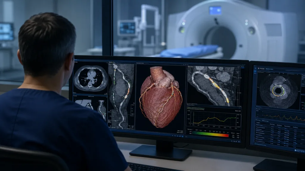
Even a sophisticated model may therefore identify statistical vulnerability without being able to say with certainty which plaque will cause harm. The distinction between recognizing features associated with previous heart attacks and prospectively predicting future events will be central to how the AI-PREDICT findings are eventually interpreted.
Can the AI-PREDICT retrospective design produce evidence strong enough for clinical adoption?
The planned size and international scope give AI-PREDICT the potential to become a meaningful cardiovascular imaging dataset. Approximately 1,000 subjects across more than 20 sites could provide a diverse range of scanners, imaging protocols, populations, clinical pathways, and disease presentations. Central analysis by a core laboratory blinded to outcomes should reduce the risk that knowledge of the heart attack influences how plaque features are measured.
The design also includes two useful comparison levels. Investigators can compare the culprit lesion with bystander lesions in the same myocardial infarction patient, then compare those findings with lesions in matched controls who did not experience an event. If the same features remain informative across both comparisons, the study would offer a stronger argument that the signal is linked to lesion behavior rather than merely reflecting a generally sicker patient.
Retrospective evidence nevertheless has unavoidable limitations. Eligible myocardial infarctions may have occurred as long as 36 months after the baseline CCTA, leaving time for plaques to progress, regress, remodel, or change composition. The lesion identified as responsible for the event must be matched back to an earlier scan, and that adjudication can become difficult when coronary anatomy is complex, medical records are incomplete, or several lesions are plausible candidates.
Image quality is another concern. Real-world CCTA data vary because of heart rate, motion, calcification, contrast timing, radiation settings, scanner generation, reconstruction methods, and local acquisition practices. International heterogeneity can strengthen generalizability, but it can also introduce noise and expose performance differences that are not apparent in tightly controlled development datasets.
The announcement does not establish the complete statistical analysis plan, exact primary endpoint, prespecified performance thresholds, or rules for handling missing and non-diagnostic scans. Those details will matter as much as the final sample size. Clinicians and regulators will need to see whether any lesion-level model adds discrimination, calibration, and clinical value beyond established information such as stenosis severity, total plaque burden, calcium, conventional risk scores, and functional assessment.
The definition of event-free controls will also require scrutiny. A control who has not experienced an event during the available follow-up period may still have clinically significant disease or suffer an event later. Differences in follow-up duration, medication use, interventions, and access to healthcare could affect comparisons unless they are adequately addressed.
Why combining PlaqueIQ with FFR-CT could sharpen Elucid’s competitive position
Elucid is entering a competitive field in which CCTA is becoming a platform for layered coronary assessment rather than a single anatomical test. HeartFlow offers non-invasive anatomical, physiological, and plaque analysis, while Cleerly markets artificial intelligence tools for plaque characterization, stenosis, and ischemia assessment. The commercial contest is increasingly about which platform can combine reliable measurements, clinically useful reporting, reimbursement, workflow compatibility, and evidence that changes decisions.
PlaqueIQ gives Elucid a cleared foundation in plaque quantification and morphology. Its FFR-CT software is intended to estimate the physiological effect of coronary disease without an invasive pressure-wire procedure, but the product remains under FDA review. AI-PREDICT is expected to use both technologies, allowing investigators to examine whether plaque biology and physiology are more informative together than either is alone.
That combination is strategically important because a lesion can look biologically concerning without causing major flow limitation, while another lesion may impair blood flow without showing the same plaque composition. A platform that integrates both dimensions may offer a more complete explanation of present disease and future risk.
The commercial value will depend on whether the combined output helps clinicians make a clearer decision rather than simply adding more measurements to an already complex report. Cardiology and radiology teams already receive information on stenosis, vessel location, plaque burden, calcium, symptoms, functional significance, and broader clinical risk. Lesion-level artificial intelligence must simplify or improve decision-making, not produce another layer of data that requires extensive interpretation.
Elucid must also remain precise about the boundary between its cleared and investigational capabilities. PlaqueIQ is cleared as an image analysis aid for plaque assessment. FFR-CT and stenosis functions remain under review, and AI-PREDICT is research rather than proof that the platform can prospectively prevent myocardial infarction. Strong results could support future regulatory discussions or product development, but they would not automatically expand the existing indication for use.
What hospitals and imaging programs need before lesion-level artificial intelligence becomes routine
Clinical adoption will require more than a high area under the curve or a statistically significant association. Hospitals will need to understand how the analysis fits into CCTA ordering, image transfer, reporting, cardiology review, radiology review, treatment planning, and follow-up. Turnaround time, failure rates, quality control, cybersecurity, data residency, and integration with picture archiving and electronic health record systems can determine whether an advanced algorithm becomes routine or remains limited to specialist centers.
The FDA clearance summary for PlaqueIQ described a cloud-based workflow in which Elucid analysts generate segmentations and apply plaque algorithms before results are returned to physicians. That service model may improve standardization and reduce the need for every hospital to train its own advanced segmentation team. It also creates operational dependencies involving image upload, analyst capacity, turnaround consistency, rework procedures, and cross-border handling of health data.
Scalability will become more important if lesion-level analysis generates wider interest in quantitative CCTA. A specialist core laboratory can support research and early commercial use, but routine deployment across large health systems requires predictable processing capacity. Hospitals are unlikely to redesign clinical pathways around a product if reporting times fluctuate or a substantial proportion of scans cannot be analyzed.
Reimbursement conditions have become more supportive for quantitative coronary plaque analysis, and coverage decisions have expanded access to advanced CCTA services. Even so, payment does not guarantee adoption. Procurement teams will compare the cost of analysis with its effect on downstream testing, invasive angiography, medication decisions, repeat imaging, physician time, and patient outcomes.
A platform that identifies more abnormalities without reducing uncertainty may increase expenditure rather than create value. Elucid will therefore need evidence showing not only that its measurements are accurate, but also that the information changes management efficiently and avoids unnecessary procedures.
Training and communication will be equally important. Lesion-level risk scores require clinicians to understand what the score measures, how uncertainty is presented, when image quality is insufficient, and how findings should be reconciled with symptoms and established clinical tests. Hospitals will also require clear governance around algorithm updates, performance monitoring, subgroup bias, and responsibility when software output conflicts with physician interpretation.
Could highly specific plaque predictions improve prevention without encouraging overtreatment?
The long-term appeal of lesion-level analysis is the possibility of moving from broad risk categories toward more individualized prevention. A patient with extensive but apparently stable disease may require a different strategy from a patient whose imaging shows a smaller number of biologically concerning lesions. More precise characterization could also help research programs select higher-risk populations for trials and evaluate whether therapies change plaque composition over time.
The unresolved problem is actionability. Identifying a lesion that appears vulnerable does not establish that locally treating that lesion will prevent a heart attack, particularly when it is not severely obstructive. Coronary events arise from a systemic disease process, and the plaque that looks most threatening on one scan may not be the lesion that later causes the event.
Intervening on imaging findings without validated treatment pathways could expose patients to unnecessary invasive procedures, repeated testing, or intensified therapy without proven net benefit. It could also encourage false reassurance when a scan does not identify a clearly dominant high-risk lesion, even though cardiovascular risk has not disappeared.
For that reason, AI-PREDICT will be most persuasive if it shows incremental value over patient-level and vessel-level measures, not merely an association with past events. The field will need thresholds that are reproducible, interpretable, and linked to a defined clinical response. A model that ranks lesions but leaves physicians uncertain about what to do next may be scientifically interesting while remaining commercially and clinically difficult to implement.
Prospective validation will eventually be necessary. A future study would need to apply the model before events occur, test it across representative populations, measure how often it changes management, and determine whether those changes improve outcomes without creating excessive downstream procedures. That is a much higher evidence bar than retrospective classification, but it is the standard required for a tool that aspires to guide prevention.
Which results will determine whether AI-PREDICT becomes more than a large imaging database?
The next important disclosures will involve study execution rather than enrollment headlines. Industry observers will look for the speed at which the remaining sites begin contributing cases, the proportion of scans that pass quality review, the demographic and geographic diversity of the dataset, and the consistency of lesion measurements across scanner types and clinical settings.
The final analysis will need to show how PlaqueIQ and FFR-CT perform separately and together. It should also explain whether the model remains useful after accounting for stenosis, calcium, total plaque burden, clinical risk, and other established predictors. A modest improvement over existing assessment methods may not be enough to justify the added cost and workflow requirements.
Subgroup performance by sex, age, ethnicity, diabetes status, disease severity, scanner platform, and time from CCTA to myocardial infarction will be critical for judging whether the results are broadly transportable. Artificial intelligence models can appear accurate at the population level while performing less reliably in smaller or underrepresented groups, making detailed subgroup reporting essential.
AI-PREDICT is therefore best viewed as an evidence-building study for Elucid’s lesion-centric cardiovascular imaging strategy, not as validation of a ready-made heart attack prediction tool. Its most valuable contribution may be the creation of a rigorously analyzed dataset that clarifies which plaque features deserve prospective testing. The commercial opportunity becomes larger if those features can be converted into a reproducible report, an actionable treatment pathway, and eventually proof that acting on the information improves patient outcomes.
For Elucid, the study connects an FDA-cleared plaque analysis product with a wider platform ambition that includes anatomy, morphology, and physiology. Success will depend less on whether the software can find differences between culprit and non-culprit plaques after an event, and more on whether those differences can be recognized early enough, reliably enough, and usefully enough to change what clinicians do before the event occurs.
Study design, regulatory status, reimbursement context, and competitive positioning were verified using the announcement, FDA records, current company materials, and official industry sources.