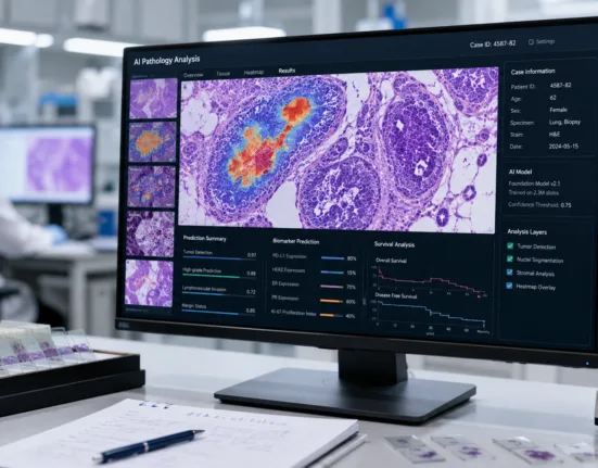
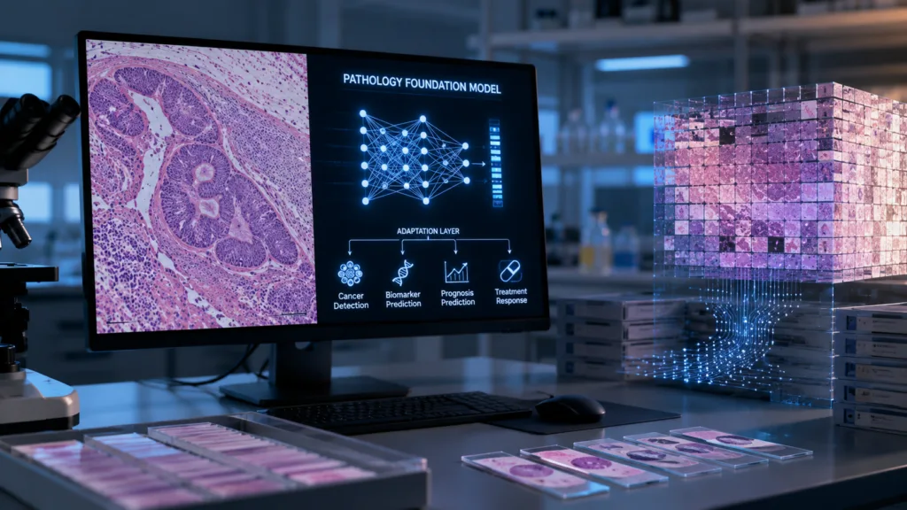
Traditional medical AI is usually built around one narrowly defined problem. Developers collect labeled breast pathology slides, train an algorithm to recognize a particular feature and validate that model specifically for breast pathology. A different cancer, biomarker or scanner environment may require another dataset and another model. Foundation models attempt to reverse that development architecture by training one very large model on vast collections of pathology images so it learns general representations of cells, tissue architecture and disease patterns that can subsequently be adapted to many clinical tasks with much smaller amounts of labeled data.
The scale is growing rapidly. A 2026 Nature Medicine study described PRISM2, a multimodal pathology foundation model trained using 2.3 million whole-slide images and 14 million question-and-answer pairs derived from approximately 700,000 pathology reports. The system was designed not merely to encode image patches but to connect whole-slide morphology with diagnostic language and reasoning, achieving or exceeding the balanced accuracy of calibrated clinical-grade products in prompt-based cancer-detection evaluations involving prostate, breast and breast lymph-node pathology.
Why is a pathology slide unusually difficult for artificial intelligence?
A digitized pathology slide is not an ordinary photograph. Whole-slide images can contain billions of pixels because a pathologist may need to move from a low-magnification view of tissue architecture to microscopic examination of individual cells. The clinically relevant abnormality may occupy only a tiny fraction of that enormous image.
This creates a computational hierarchy. AI must understand cells, groups of cells, tumor boundaries, stroma, necrosis, inflammatory infiltrates and the relationship between geographically separated regions of tissue. A model trained only on isolated image patches can learn useful local patterns while missing the case-level relationships a pathologist uses to reach a final diagnosis.
Human pathology is also combinatorial rather than object-based. A pathologist rarely identifies disease by spotting one equivalent of a cat or traffic sign. Diagnosis emerges from combinations of nuclear appearance, tissue organization, cellular proportions, anatomical context and sometimes multiple slides stained using different techniques.
This is one reason researchers have cautioned against assuming that architectures successful on ordinary photographs will transfer naturally into medicine. A 2026 Nature Biomedical Engineering commentary argued that tissue pathology violates several assumptions underlying general vision models and may require architectures built more explicitly around biological complexity.
What does a foundation model actually learn from millions of pathology images?
Most foundation models initially learn representations rather than a final diagnosis. During training, they discover visual relationships that allow similar cellular and tissue structures to be represented mathematically near each other even when the model has not been provided with exhaustive human labels for every feature.
Those learned representations can then support downstream tasks. A relatively small labeled dataset can teach another model to use the foundation representation for tumor detection, subtyping, biomarker prediction, survival modeling or other questions, potentially requiring far less task-specific annotation than training an entire diagnostic algorithm from scratch.
The earlier Virchow pathology foundation model demonstrated this promise across common and rare cancers. Researchers reported a specimen-level area under the receiver operating characteristic curve of 0.95 for pan-cancer detection across nine common and seven rare cancers, and models built on Virchow could achieve performance comparable with some tissue-specific clinical-grade systems using less training data.
That is particularly relevant for rare cancers, where collecting tens of thousands of expertly labeled cases may be impossible. A broadly trained model that already understands basic tissue morphology can potentially be adapted using far smaller rare-disease datasets.
Why are researchers now adding pathology reports and language to image models?
A pathology report represents compressed expert reasoning. It does not merely say that a slide contains pink and purple shapes; it describes diagnosis, grade, tissue relationships, biomarkers and other clinically meaningful conclusions derived from those visual patterns.
Multimodal models attempt to align images with that language so the AI learns not just whether two patches look similar but how morphological features relate to concepts used by pathologists. PRISM2 was trained using millions of whole-slide images together with question-and-answer data derived from hundreds of thousands of reports, allowing researchers to query the system using language rather than building a separate model for every diagnostic prompt.
This creates the possibility of a pathology assistant capable of answering several questions about one specimen: whether tumor is present, what histological subtype is likely, which regions deserve closer attention and potentially whether visible morphology contains information associated with molecular biomarkers.
It also increases the risk of plausible but incorrect answers. A model capable of producing fluent diagnostic language may appear more confident than its underlying evidence justifies, making clinical validation and human oversight essential.
Can AI predict molecular biomarkers from an ordinary H&E slide?
Sometimes, at least statistically. Genetic alterations can change cellular morphology and the tumor microenvironment in ways that are visible microscopically even when the underlying mutation itself cannot be seen directly. Machine-learning systems can exploit patterns distributed across thousands of cells that may be too subtle or inconsistent for humans to use reliably.
Foundation models have consequently been investigated for predicting molecular characteristics, treatment-relevant biomarkers and patient prognosis directly from routine hematoxylin-and-eosin slides.
That does not mean histology AI can automatically replace molecular testing. A model prediction is an inferred probability based on morphological correlates, whereas sequencing or a validated immunohistochemistry assay may measure the biomarker more directly. The acceptable role will depend on performance: AI might become a triage tool determining which samples need expensive molecular testing, or in specific circumstances it could eventually become a validated diagnostic method itself.
The economic opportunity is substantial because H&E slides already exist for enormous numbers of cancer patients. Extracting additional information from an image already produced during routine diagnosis requires no new tissue collection, which resembles the value proposition driving opportunistic analysis in medical imaging.
What is the hidden danger of scanner and laboratory bias?
Pathology slides differ for reasons unrelated to biology. Tissue fixation, section thickness, staining chemistry, laboratory workflow, scanner manufacturer and image-compression settings can all change the visual appearance of a digital slide.
An AI system can accidentally learn those technical signatures. If a training hospital happens to process aggressive cancer cases differently from routine cases, the model may discover the laboratory workflow rather than the cancer biology and still appear highly accurate during internal testing.
Research published in Nature Communications in 2026 showed that pathology foundation models remain vulnerable to non-biological confounders associated with laboratory procedures and scanner hardware, creating a serious challenge for generalization across institutions.
This is why external validation matters enormously. An algorithm that performs beautifully on another random subset of slides from the same institution may fail once it encounters tissue processed in another country, stained with different reagents or digitized using another scanner.
Does a foundation model need FDA clearance before a pathologist can use it clinically?
The foundation model itself can be a research platform rather than a medical device. Regulatory requirements depend on what the deployed software is intended to do and how its output influences patient care.
A model used to generate research embeddings has a very different regulatory profile from software marketed to detect prostate cancer, quantify a treatment biomarker or issue a diagnostic recommendation. Clinical products built on foundation models therefore still need appropriate validation and regulatory pathways for their intended use rather than inheriting authorization because the underlying general model performed well in academic benchmarks.
This also complicates continuous model updates. If a foundation model is retrained on substantially more data or its architecture changes, developers need a regulatory strategy ensuring that improvements in one area have not degraded performance in previously validated patient populations.
Could one pathology foundation model eventually replace dozens of individual AI products?
Technically, that is the ambition. Economically, it could be transformative because hospitals do not want separate disconnected applications for every tumor type and biomarker. A common pathology intelligence layer that supports many validated clinical tasks could simplify deployment, integration and maintenance.
The emerging evidence suggests that foundation models can provide generalizable representations across many tissues, and new systems such as PRISM2 are beginning to operate at the whole-slide and multimodal level rather than merely encoding small tiles. At the same time, a 2026 benchmark comparing 32 foundation models emphasized that performance and generalizability remain variable across models, datasets and tasks, meaning scale alone does not guarantee clinical robustness.
The more realistic future may therefore separate the platform from the regulated application. One enormous foundation model can provide the underlying representation, while individual validated layers translate that intelligence into specific clinical claims such as cancer detection, biomarker scoring or prognostic estimation.
The decisive question is not whether an AI system can learn from millions of slides. Researchers have already shown that it can. The test is whether it can recognize cancer biology rather than laboratory fingerprints, maintain performance when transferred between hospitals and provide clinically useful outputs reliably enough for a pathologist to trust when one incorrect interpretation concerns a real patient rather than a benchmark score.