Northwell Health’s Feinstein Institutes for Medical Research has released what it describes as the world’s first comprehensive anatomical map of the human vagus nerve, making multimodal data from 30 human donors and 60 vagus nerves available to researchers through the National Institutes of Health-funded SPARC platform. The resource combines anatomical dissection, microcomputed tomography, ultrasound and immunohistochemistry to examine how nerve bundles and individual fiber types are organised along the nerve.
The announcement represents a research infrastructure milestone rather than a clinical trial result, regulatory decision or demonstration that a particular neuromodulation therapy improves patient outcomes. Its immediate value lies in giving scientists and device developers a substantially larger human anatomical reference against which they can test electrode designs, computational models, stimulation strategies and assumptions previously derived from small studies or animal anatomy.
The dataset was developed through the Reconstructing Vagal Anatomy project, known as REVA, following a multiyear National Institutes of Health award announced in 2022. The programme was created to address a persistent problem in bioelectronic medicine: researchers understand many of the physiological functions influenced by the vagus nerve, but still have an incomplete picture of how sensory and motor fibers are arranged within its internal fascicles and how those fascicles change as the nerve travels from the brainstem towards the chest and abdomen.
What exactly has the Feinstein Institutes released through the REVA vagus nerve project?
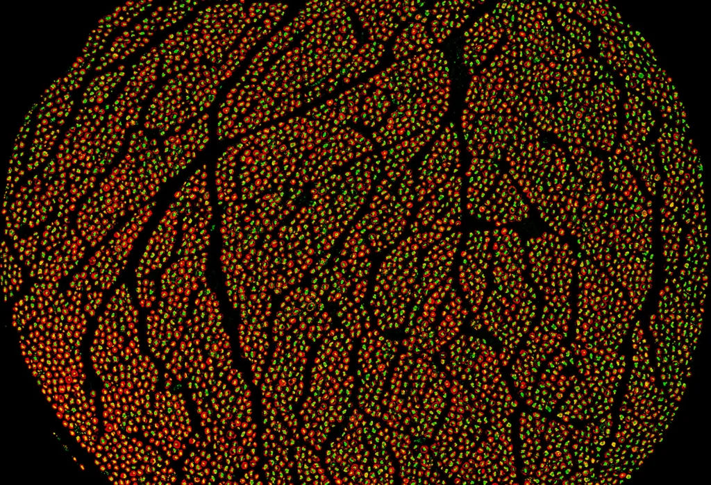
The vagus nerve map is not a single conventional anatomical illustration. It is a collection of large, donor-level datasets containing photographs and videos from anatomical dissections, ultrasound recordings, high-resolution microCT scans and microscopic images produced with histological and immunohistochemical staining.
Published SPARC records show that individual donor datasets can contain hundreds of files and approach two terabytes in size. These records describe nerves dissected from the upper cervical region through the thorax and abdomen, with branches identified according to the organs or tissues they appeared to reach. Nerve segments were subsequently imaged and stained for markers including neurofilament, myelin basic protein, choline acetyltransferase and, in some samples, tyrosine hydroxylase.
MicroCT provides a three-dimensional view of the fascicles contained within the vagus nerve, while immunohistochemistry can help distinguish structural and molecular features of different fibers. Ultrasound adds a modality more closely connected with how the nerve may appear during a clinical or surgical procedure. Cross-registering these layers could eventually allow researchers to move between macroscopic anatomy, fascicular organisation and microscopic fiber characteristics within the same donor.
That multimodal structure is arguably more consequential than the headline claim of being first. A map built from only one imaging method may show where a fascicle is located without revealing which fiber populations it contains. Combining modalities offers a path towards richer computational models, although the usefulness of those models will depend on the completeness and consistency of the underlying annotations.
Why has incomplete vagus nerve anatomy limited more selective stimulation strategies?
Existing vagus nerve stimulation systems generally deliver electrical energy to a nerve containing many fiber populations associated with different organs and physiological functions. Depending on the device, indication, electrode configuration and stimulation settings, that can make selective recruitment difficult.
The human vagus is not a neat cable in which every organ-specific pathway remains isolated from the brainstem to its destination. Fascicles divide, merge and change position along the nerve. A previous microCT study of human cervical vagus nerves found a fascicle splitting or merging event approximately every 560 micrometres within the examined surgical window. Other research has identified substantial differences in fascicle number, cross-sectional area, fiber composition and morphology between individuals and between the right and left nerves.
A peer-reviewed anatomical study published in 2026 traced cardiac, pulmonary and recurrent laryngeal branches in human cadaveric nerves. It found that some organ-associated fascicles retained partial organisation near their branch entry points but subsequently merged as they travelled through the nerve. The study also reported differences between left and right vagus nerves, reinforcing the possibility that a stimulation strategy optimised for one anatomical pattern may not perform identically in another person.
These findings explain why precision neuromodulation is not simply a matter of shrinking an electrode. A smaller contact may still stimulate an unintended fiber population when fascicles are densely packed, continuously reorganising or composed of mixed functional fibers. The challenge is therefore anatomical, electrical and computational at the same time.
How could open human vagus nerve data change neuromodulation device development?
The most immediate beneficiaries are likely to be researchers building computational models of nerve stimulation. Device teams can use reconstructed fascicle geometries to estimate which fibers may be recruited at different electrode positions, current amplitudes, pulse widths and stimulation patterns before committing to expensive prototypes or clinical studies.
Human anatomical datasets may also improve the translation of findings from animal models. Animal studies have provided evidence that vagal fibers can show organ-specific and function-specific organisation, but anatomical differences between species complicate direct application to human devices. A larger human reference library allows developers to test whether a design remains selective across multiple nerve shapes rather than performing well only on one idealised model.
Data volume creates its own engineering challenge. Manually segmenting every fascicle across thousands of microCT slices is slow and vulnerable to observer variation. Researchers have therefore been developing deep-learning systems to automate the three-dimensional segmentation of the epineurium and internal fascicles. One earlier study reported that an automated model processed hundreds of images in seconds compared with dozens of hours for manual segmentation, although it could under-detect smaller fascicles. A more recent anatomy-aware approach has sought to improve segmentation while preserving nerve topology.
The open-data model could also change the competitive structure of bioelectronic medicine. Basic anatomy may increasingly become shared infrastructure, while differentiation moves towards electrode geometry, implantation methods, stimulation algorithms, sensing capabilities, closed-loop control and clinical evidence. Companies may no longer need to build every anatomical reference from scratch, but they will still need to show that their specific device can target the intended pathway reproducibly and safely.
Why should the vagus nerve map not be treated as clinical validation for new therapies?
Cadaveric anatomy can show the location, shape and composition of nerve structures, but it cannot independently demonstrate how those structures behave in a living person during electrical stimulation. It also cannot establish that activating a particular region will improve a disease outcome.
Tissue fixation, embalming, dissection, staining and imaging may alter dimensions or introduce artefacts. Donor age, sex, medical history and anatomical variation may influence whether the available specimens represent the population intended to receive a future device. Functional identity can also become uncertain after fascicles merge, particularly when imaging resolution can follow the fascicle boundary but not the trajectory of every individual axon.
Several SPARC donor records describe the current material as an initial or incomplete release, with additional processed data, derivative outputs or protocols expected. The word “comprehensive” is therefore best interpreted as the breadth of the project’s anatomical ambition and multimodal collection, not as evidence that every analytical layer has been completed or clinically validated.
Researchers will need to connect the anatomical data with electrophysiology, physiological responses, intraoperative measurements and prospective human studies. A stimulation contact predicted to reach cardiac-associated fibers, for example, must still be tested to determine whether it produces the expected cardiovascular effect without unacceptable laryngeal, pulmonary or gastrointestinal consequences.
What does the SetPoint rheumatoid arthritis approval reveal about the commercial pathway?
The clinical and regulatory potential of vagus nerve stimulation is no longer confined to neurological indications. In July 2025, the United States Food and Drug Administration approved the SetPoint System for adults with moderately to severely active rheumatoid arthritis who had an inadequate response, loss of response or intolerance to at least one biological or targeted synthetic disease-modifying antirheumatic drug.
That approval is significant because it demonstrates a regulatory pathway for an implanted vagus nerve stimulator intended to influence an immune-mediated disease. It does not, however, mean that a general anatomical map automatically de-risks other inflammatory, cardiovascular, metabolic or gastrointestinal indications.
The SetPoint System underwent device-specific clinical evaluation and remains subject to post-approval surveillance. The FDA’s post-approval requirements include monitoring implantation-related events, device performance, stimulation-related safety and clinical outcomes in routine practice. The regulator expects approximately 150 participants in a prospective multicentre observational registry, illustrating how evidence generation continues after market authorisation.
For future developers, the lesson is clear. Better anatomical targeting may strengthen device design and support more rational selection of stimulation parameters, but regulators will still require evidence tied to the final hardware, software, implantation procedure, intended patient population and proposed clinical claim.
What must happen before the new vagus nerve map changes routine patient care?
The first test will be whether independent researchers can reproduce the project’s segmentations, compare donors consistently and convert the raw data into models that predict stimulation responses accurately. Open access helps, but very large datasets require substantial storage, computing capability and specialised expertise, potentially limiting immediate use to well-resourced academic centres and device companies.
The second test involves population coverage. Developers will need to understand how vagal anatomy varies with sex, age, body size, disease, previous surgery and other clinically relevant factors. A population atlas may ultimately be more useful than a single averaged nerve because an average geometry can hide the very anatomical differences that cause variable stimulation responses.
The third test is functional validation. Anatomical labels must be linked to measurable physiological endpoints in living humans. That may require imaging, nerve recordings, evoked responses, carefully controlled stimulation studies and, where justified, clinical trials examining whether greater selectivity leads to better outcomes or fewer adverse effects.
The Feinstein Institutes release gives bioelectronic medicine a richer anatomical foundation and may reduce duplicated work across the field. Its longer-term impact will depend on whether researchers can turn a complex library of cadaveric images into validated, patient-relevant rules for placing electrodes and selecting stimulation parameters. The map makes the nerve more visible. The harder task is proving that this visibility can make neuromodulation more predictable.