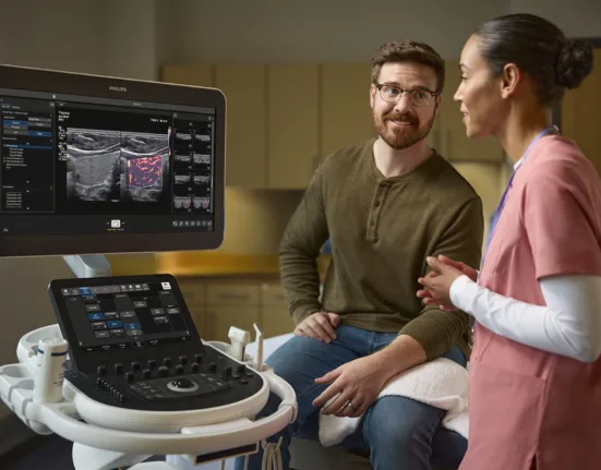
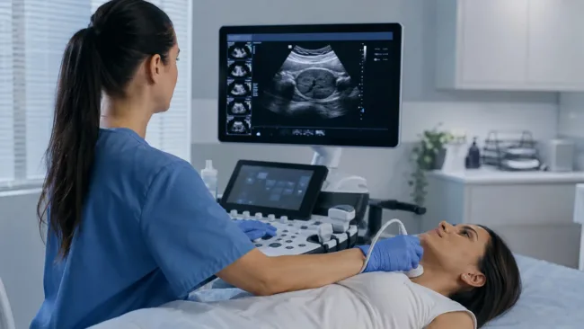
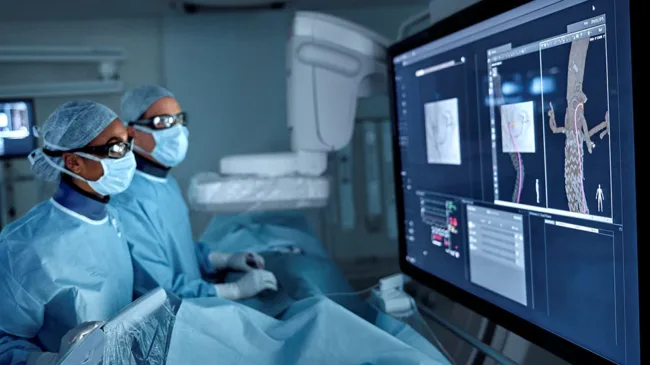
Catheter ablation has become one of cardiology’s most sophisticated minimally invasive treatments, yet physicians performing the procedure still face a basic imaging limitation: they can navigate the heart with electrical maps and X-ray fluoroscopy but cannot routinely see the soft tissue response to treatment with anything approaching the detail available from magnetic resonance imaging.
That gap is beginning to narrow. Royal Philips and Imricor Medical Systems, Inc. have launched an interventional magnetic-resonance laboratory configuration combining Philips’ 1.5-tesla MRI technology with Imricor’s MRI-compatible electrophysiology mapping, recording and catheter systems. The platform is intended to allow clinicians to visualize cardiac anatomy, identify arrhythmia substrate, track catheters, create electrical maps, assess lesion formation and evaluate treatment response without relying on conventional ionizing-radiation imaging as the primary environment.
The development does not mean every electrophysiology laboratory is about to replace fluoroscopy with an MRI scanner. Real-time MRI-guided intervention remains an emerging field with major equipment, workflow, device-compatibility and training challenges. Recent clinical studies, however, are beginning to show that the concept can move beyond experimental imaging and into actual catheter procedures.
Why is conventional catheter ablation partly an imaging problem?
During cardiac ablation, physicians navigate catheters through blood vessels and into specific regions of the heart responsible for an abnormal rhythm. Radiofrequency energy, cryothermal energy or newer nonthermal technologies can then modify targeted tissue so that unwanted electrical signals can no longer propagate through the same pathway.
Electroanatomical mapping systems have dramatically reduced dependence on fluoroscopy by reconstructing chamber geometry and displaying electrical activity in three dimensions. Yet these maps are models derived from catheter location and electrical measurements rather than direct, continuous visualization of the tissue itself.
Fluoroscopy presents a different limitation. X-rays are excellent for showing radiopaque catheters and devices but provide relatively poor intrinsic soft-tissue contrast. Physicians can see where an instrument is positioned, but they cannot directly observe myocardial scar, edema or lesion maturation with the same tissue detail offered by MRI.
Cardiac MRI can characterize anatomy and tissue composition, including fibrosis and scar, which is why it is already valuable before and after selected complex electrophysiology procedures. The long-term ambition of interventional MRI is to bring that tissue information into the procedure itself.
How would an interventional MRI cardiac lab actually work?
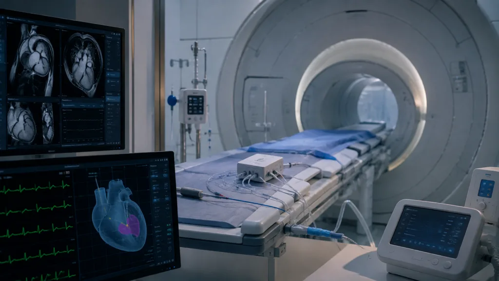
The Philips and Imricor configuration places the intervention within a 1.5-tesla MRI environment rather than attempting to move a patient back and forth between a conventional catheter laboratory and a diagnostic scanner.
Philips contributes its cardiac MRI platform, Cardiac MR Suite and SmartHeart artificial-intelligence-enabled planning tools. Imricor supplies the NorthStar interventional MRI mapping and guidance system, Advantage-MR electrophysiology recorder and stimulator and MRI-compatible Vision-MR catheter family.
Together, the technologies are intended to support active catheter tracking and electroanatomical mapping while MRI provides anatomical and tissue information. In principle, this means physicians can combine what the heart looks like, where the catheter is located and what the tissue is doing electrically within the same procedural environment.
That integration is much harder than placing ordinary electrophysiology equipment near an MRI scanner. Strong magnetic fields and radiofrequency energy can interfere with conventional cables, sensors and metallic components, while some conductive structures can heat dangerously during imaging.
Every instrument entering the room therefore becomes part of the MRI-safety problem.
Why is eliminating radiation important if modern ablation already uses less fluoroscopy?
Electrophysiology laboratories have made major progress in reducing radiation exposure through advanced mapping systems and low-fluoroscopy workflows. Some procedures can already be completed with little or no fluoroscopy by experienced teams.
The remaining issue is cumulative exposure. Electrophysiologists, nurses and technicians may participate in thousands of procedures over a career, while patients with complex arrhythmias can undergo multiple interventions.
Radiation protection also influences the ergonomics of the laboratory because staff traditionally wear heavy lead garments during fluoroscopic procedures. Reducing dependence on X-rays therefore has implications for both radiation exposure and occupational strain.
MRI creates another benefit that low-fluoroscopy mapping alone cannot provide: direct soft-tissue visualization. The strongest argument for interventional MRI may consequently be not that it removes X-rays, but that it replaces them with an imaging modality capable of showing more biologically relevant information.
Could MRI allow physicians to see whether an ablation lesion actually worked?
This is one of the most compelling possibilities.
Ablation succeeds only if the energy delivered to tissue creates a durable lesion in the correct location. Physicians currently infer lesion quality from variables including catheter contact, energy delivery, impedance changes, temperature, electrical response and mapping data.
MRI has the potential to visualize tissue changes associated with ablation, providing another way to determine whether the intended substrate has been adequately treated before the patient leaves the procedure room. Philips and Imricor specifically describe lesion assessment and evaluation of treatment response as functions their integrated environment is designed to support.
If validated clinically, this could change the procedural endpoint. Instead of concluding that an ablation is complete because the electrical signals and procedural parameters look appropriate, physicians could potentially combine electrical evidence with direct tissue imaging.
The implication is particularly important for recurrence. Arrhythmias can return when ablation lines contain gaps or when apparently treated tissue recovers electrical conduction. Imaging that helps identify incomplete lesions during the original procedure could theoretically reduce the need for repeat intervention, although large comparative trials would be required to establish that benefit.
What does the early clinical evidence show?
A 2026 Heart Rhythm report describing early clinical use of interventional cardiac MRI-guided atrial flutter ablation highlighted the ability to perform catheter ablation without ionizing radiation while obtaining real-time soft-tissue visualization. The authors focused on the technical evolution needed to translate the approach into actual procedures rather than merely demonstrating MRI compatibility in a laboratory.
Researchers have also reported a first-in-human real-time MRI-guided radiofrequency ventricular ablation procedure for idiopathic outflow-tract premature ventricular complexes. That case formed part of the VISABL-VT clinical program and used a 1.5-tesla MRI environment with dedicated MRI-compatible electrophysiology equipment and real-time catheter tracking.
Evidence from a broader 2026 meta-analysis also supports the value of incorporating cardiac MRI information into ventricular-arrhythmia ablation even when the entire procedure is not performed inside MRI. Across 18 studies involving 1,243 participants, MRI-guided approaches integrated with three-dimensional electroanatomical mapping were associated with lower ventricular-arrhythmia recurrence and higher procedural success than electroanatomical mapping alone, although the evidence combined different imaging strategies and does not prove that fully real-time MRI intervention will reproduce the same benefit.
These studies collectively show momentum, but they should not be interpreted as evidence that interventional MRI has already replaced conventional electrophysiology. The field remains early enough that technical feasibility, workflow and procedural learning remain important research questions.
Why is building an MRI-compatible catheter so difficult?
An electrophysiology catheter contains components that would be relatively unremarkable in a conventional operating environment but become challenging inside an MRI scanner.
Conductive wires can interact with the scanner’s radiofrequency fields and behave like antennas, potentially producing localized heating. Electronic components must function without creating unacceptable imaging artifacts or being disrupted by the magnetic environment.
Research published in Medical Physics in August 2026 illustrates how actively this engineering problem is being studied. Investigators developing an MRI-guided magnetically actuated robotic catheter focused specifically on mitigating radiofrequency-induced and resistive heating, underscoring that thermal safety is a fundamental requirement for future MRI-compatible interventional robotics.
The challenge extends to every accessory surrounding the procedure. Recording systems, monitoring equipment, defibrillation capability, pumps, displays and emergency tools all need workflows that remain safe inside or near the magnetic field.
That systems-level complexity is one reason interventional MRI has taken much longer to mature than diagnostic cardiac MRI.
Could artificial intelligence make MRI-guided intervention easier?
Artificial intelligence could become an important enabling layer because MRI generates large volumes of imaging information while electrophysiology simultaneously produces complex electrical data.
A future system could automatically segment cardiac chambers, identify scar, register images with catheter position, detect potential treatment gaps and compare pre-ablation and post-ablation tissue changes. Philips’ current configuration already incorporates SmartHeart AI-assisted cardiac planning, suggesting that automation is likely to be embedded increasingly deeply into interventional MRI workflows.
AI could also address one practical problem: cognitive overload. Giving a physician more imaging is not automatically beneficial if the additional information becomes too difficult to interpret while manipulating a catheter inside a beating heart.
The most useful systems may therefore be those that transform MRI data into a small number of clinically actionable overlays rather than simply filling the room with additional images.
Will hospitals really build dedicated MRI procedure rooms?
Infrastructure may be one of the largest barriers to widespread adoption. MRI installations require substantial capital investment, magnetic-field safety controls and specialized facility design, while electrophysiology laboratories are already expensive environments.
Philips and Imricor argue that the objective is not to perform interventions inside ordinary diagnostic MRI suites but to create dedicated interventional MRI laboratories designed around procedures. Their first configuration uses Philips’ BlueSeal magnet architecture, which contains a small sealed helium volume and is intended to simplify some siting requirements compared with conventional helium-intensive MRI systems.
Even with easier installation, hospitals will need compelling utilization economics. A dedicated iMR laboratory becomes more attractive if it can support numerous procedures across cardiology and eventually other specialties rather than being used for a narrow group of arrhythmia cases.
Philips has already signaled that cardiac electrophysiology is intended as a starting point rather than the final scope of the platform.
Could MRI guidance eventually extend beyond cardiac ablation?
The broader future of interventional MRI could include structural heart procedures, catheter-based therapies, biopsies, targeted drug delivery and interventions elsewhere in the body where direct soft-tissue visualization provides an advantage.
The crucial transition is conceptual. Diagnostic imaging traditionally answers the question of what disease is present, while interventional imaging answers where a physician should act. Real-time MRI attempts to merge those roles so that the same imaging environment identifies pathology, guides treatment and assesses the biological response before the procedure ends.
Robotic catheter systems could extend that model even further. If engineers can develop MRI-safe robotic devices capable of precise remote manipulation, the eventual procedure room could combine MRI tissue characterization, AI-assisted targeting and robotic catheter control in one closed-loop environment.
That future remains technically demanding and will require clinical validation well beyond today’s early experience.
What will determine whether interventional MRI becomes routine?
The technology must prove that better tissue visualization translates into outcomes that matter. Hospitals are unlikely to rebuild electrophysiology infrastructure simply because MRI produces impressive images.
The strongest case would involve evidence that MRI guidance reduces arrhythmia recurrence, improves procedural success, decreases repeat ablations or enables interventions that are difficult to perform reliably with existing mapping and fluoroscopic techniques.
Procedure duration, staffing, equipment reliability, catheter cost and patient eligibility will matter just as much. MRI also introduces contraindications and workflow restrictions that may prevent universal use.
The most likely future is therefore not the sudden disappearance of the conventional electrophysiology laboratory. MRI-guided intervention may initially develop as a specialized option for selected complex arrhythmias and institutions with sufficient cardiac-imaging expertise, gradually broadening as compatible devices and clinical evidence accumulate.
What makes the current moment important is that several pieces are finally converging. MRI scanners are becoming easier to site, dedicated interventional catheters exist, real-time tracking has reached human procedures, AI can help process increasingly complex imaging, and commercial platforms are beginning to assemble these components into one laboratory.
For decades, electrophysiologists have become extraordinarily skilled at treating cardiac tissue they can only partially visualize. Interventional MRI is built around a very different proposition: the future of cardiac ablation may be defined not only by better energy sources or smarter electrical maps, but by finally allowing physicians to see the tissue they are trying to change while they are changing it.