LUMA Vision is preparing to commercialize VERAFEYE in the United States after receiving FDA clearance that expands the system’s imaging, navigation and procedural-guidance capabilities. The Dublin-based medtech company says VERAFEYE can create live three-dimensional models of the left and right atria, continuously track imaging and ablation catheters and document treatment delivery directly on reconstructed anatomy. LUMA Vision is targeting an early-2027 commercial launch.
The clearance adds to earlier FDA authorizations covering the VERAFEYE Imaging and Guidance System and VERAFEYE Imaging Catheter. FDA records show the guidance system received a substantial-equivalence decision in June 2026, while the imaging catheter received a separate clearance in July.
Why are cardiac ablation procedures becoming an imaging problem?
Atrial fibrillation ablation requires physicians to navigate catheters inside a moving, patient-specific three-dimensional organ while avoiding structures that may be only millimetres away from intended treatment sites. Electrophysiologists therefore combine electrical mapping, fluoroscopy, intracardiac echocardiography and increasingly sophisticated software to understand catheter position and anatomy.
The challenge is that information frequently arrives from separate systems. One screen may show electrical mapping, another ultrasound and another fluoroscopic images. Operators mentally integrate these sources while simultaneously manipulating catheters and delivering ablation.
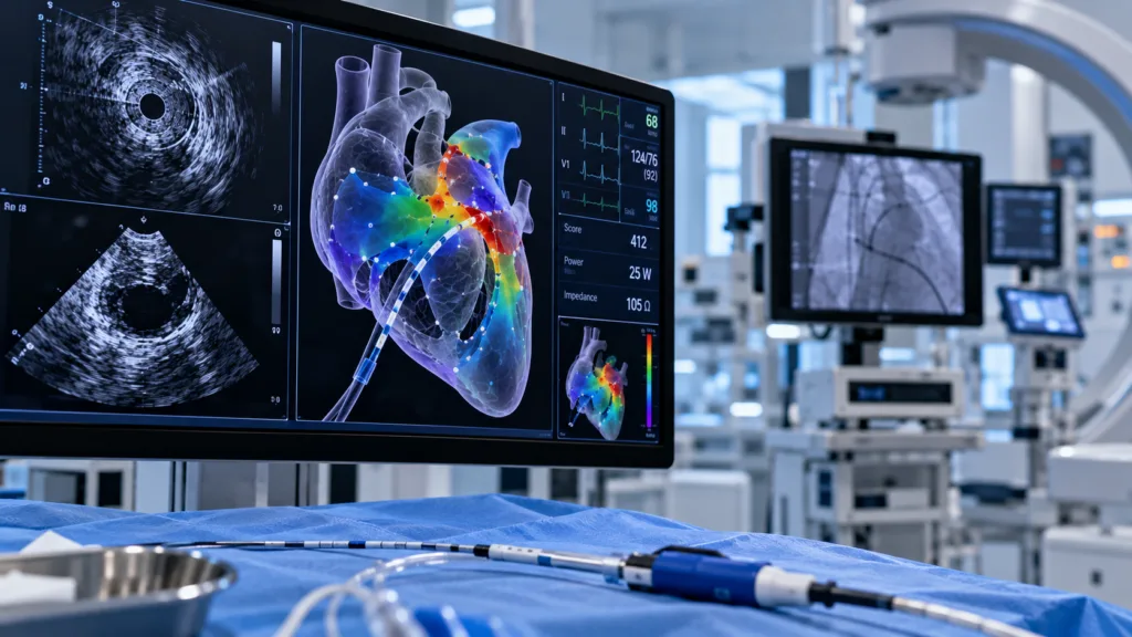
LUMA Vision is trying to collapse parts of that fragmented workflow. VERAFEYE uses a single 360-degree ultrasound imaging catheter to collect raw data and construct real-time two-dimensional and three-dimensional anatomical views, while electromagnetic tracking maintains visibility of the imaging catheter and treatment catheter.
How is VERAFEYE different from conventional intracardiac echocardiography?
Traditional intracardiac echocardiography provides ultrasound images from inside the heart and is already widely used to guide transseptal access, visualize anatomy and monitor complications. The physician usually manipulates the catheter to obtain different views and interprets two-dimensional image planes.
VERAFEYE aims to automate more of that spatial reconstruction. LUMA Vision says the system can generate three-dimensional models of the atria with CT-like anatomical accuracy while maintaining continuous visualization without requiring repeated manual repositioning of the imaging catheter.
The difference is therefore not simply image quality. The platform is designed to transform ultrasound data into an anatomical environment in which physicians can navigate, mark treatment locations and track other devices.
Why could continuous catheter tracking matter during ablation?
Catheter ablation succeeds only if energy reaches the intended tissue in a sufficiently durable pattern. Missing a region can allow electrical reconnection and recurrent arrhythmia, while unnecessary treatment increases procedural exposure.
VERAFEYE allows physicians to document ablation delivery directly on the live anatomical model and maintain simultaneous tracking of the imaging and treatment catheters.
This could be particularly useful as pulsed-field ablation accelerates procedure speed. Faster energy delivery makes efficient mapping and confirmation more important because physicians need to understand exactly where treatment has occurred even as procedural times decrease.
The strategic competition in electrophysiology is consequently shifting from individual catheters toward integrated platforms combining imaging, mapping, energy delivery and software. LUMA Vision is attempting to enter that ecosystem from the imaging-and-guidance side.
Does the platform actually use artificial intelligence?
LUMA Vision describes VERAFEYE as an AI-driven platform, but that phrase needs careful interpretation. Its immediate cleared functions involve acquisition and reconstruction of anatomy, catheter tracking, visualization and procedural guidance.
The company’s longer-term ambition goes further. As VERAFEYE creates a database of anatomical information across thousands of procedures, LUMA Vision says future guidance capabilities could increasingly help physicians decide rather than merely visualize. That future capability should not be confused with what current FDA clearance establishes.
This distinction matters throughout medical AI. Regulatory clearance often applies to specific defined functions, while companies may simultaneously describe broader future platforms that remain under development.
Can a smaller medtech company break into an EP market dominated by large platforms?
The electrophysiology market includes some of the largest medical-device companies in the world. Abbott Laboratories, Johnson & Johnson, Boston Scientific and Medtronic have extensive physician relationships, installed mapping systems and broad catheter portfolios.
LUMA Vision therefore cannot compete only by offering an interesting image. A new platform must fit existing workflows, integrate with third-party tools, justify capital expenditure and provide enough clinical or procedural improvement to convince hospitals that another system belongs in the laboratory.
Interoperability may help. LUMA Vision describes VERAFEYE as capable of tracking third-party ablation catheters rather than requiring physicians to use only one proprietary therapeutic device. That can reduce adoption resistance because hospitals do not necessarily need to abandon existing ablation preferences to use the imaging system.
Why might structural heart become an important second market?
The company says it ultimately plans to extend VERAFEYE beyond arrhythmia procedures into left atrial appendage and structural-heart interventions.
That expansion makes strategic sense because structural-heart procedures depend heavily on imaging. Transcatheter valve repair, replacement and left atrial appendage closure require continuous understanding of device position relative to complex anatomy.
Today, many structural-heart procedures rely heavily on transoesophageal echocardiography, often involving separate imaging specialists and general anaesthesia depending on the intervention. Intracardiac systems capable of producing sufficiently informative real-time 3D views could potentially change some workflows, although each application would require appropriate regulatory evidence.
A platform capable of serving both electrophysiology and structural heart would also create a larger commercial opportunity than an AFib-only imaging device.
How much has LUMA Vision invested in reaching this point?
LUMA Vision, previously known as OneProjects, raised approximately $17 million in an earlier funding round to advance its four-dimensional cardiac imaging platform. Investors included LSP, Atlantic Bridge Ventures, imec.xpand and the European Innovation Council Fund.
That venture-backed structure creates a different commercial challenge from a product launch inside a multinational medtech company. LUMA Vision must build production, clinical support, physician training and a sales organisation while competing with companies that already have thousands of employees interacting with cardiology customers.
FDA clearance substantially de-risks the technology, but commercial adoption remains capital intensive. Successful early installations will be important not only for revenue but for demonstrating that the platform deserves a permanent place in increasingly crowded electrophysiology labs.
What evidence will physicians need after launch?
Regulatory clearance establishes substantial equivalence and permits marketing for the cleared functions; it does not prove that VERAFEYE improves long-term ablation outcomes compared with established imaging strategies.
Physicians will therefore look for practical measures first. Does the system reduce imaging-catheter manipulation? Does it shorten mapping time? Can it reduce fluoroscopy? Does it improve visualization of difficult anatomy, decrease procedure duration or reduce the need for additional imaging equipment?
Longer-term studies can address harder endpoints such as complications, repeat ablation and arrhythmia recurrence. Those outcomes depend on many factors beyond imaging, making large comparative studies necessary before attributing clinical differences to the platform itself.
Is “procedural intelligence” a real category or simply branding?
The term risks becoming marketing language if it merely describes imaging plus software. LUMA Vision’s opportunity is to make it tangible by showing that integrated data materially changes physician workflow.
The category becomes more meaningful if the platform progresses from showing anatomy to contextualizing treatment, recognizing patterns and supporting decisions based on thousands of previous procedures. That would move cardiac imaging toward the same evolution occurring elsewhere in medicine, where software increasingly converts raw data into actionable procedural guidance.
Yet automation must remain carefully bounded. Electrophysiology involves complex anatomy and dynamic clinical decisions, and physicians will remain responsible for interpreting information and choosing treatment strategies.
VERAFEYE therefore enters the market at an interesting moment. Ablation energy technology is advancing rapidly, but faster therapeutic tools increase the value of knowing exactly where the catheter is, what tissue has been treated and what anatomy remains. LUMA Vision is betting that the next important EP platform will not simply deliver energy more efficiently; it will make the entire procedure easier to see and understand.